Category: Science

The scope of scientific study is vast, and it encompasses fascinating and intricate disciplines. Learn more about the most interesting topics in science.

  • Facts About Jealousy That Will Help You Better Understand Its Nature

    Facts About Jealousy That Will Help You Better Understand Its Nature

    1. Jealousy Doesn’t Necessarily Harm Relationships

    There are two primary types of this emotion:

    • Reactive jealousy arises in response to real threats—for example, if your partner is genuinely flirting with someone else. This is a normal emotional reaction.
    • Suspicious jealousy appears without objective grounds and is usually linked to low self-esteem. It can damage relationships through excessive control, pressure, and distrust. It also negatively affects mental and physical health, potentially causing headaches, stomach pain, blood pressure fluctuations, sleep disturbances, and even a weakened immune system. Over time, suspicious jealousy can develop into a chronic condition associated with anxiety disorders, depression, or obsessive-compulsive behaviors.

    While suspicious jealousy is toxic, reactive jealousy—in moderate doses—can actually be beneficial. It often signals the importance of a relationship and may prompt partners to clarify boundaries, thereby strengthening trust. People who experience reactive jealousy typically feel a strong emotional attachment and strive to maintain closeness. In such cases, jealousy isn’t a sign of weakness or dysfunction but a natural part of emotional connection. The key is to channel it constructively: replace accusations with openness, and restrictions with shared plans.

    2. Men Tend to Be More Upset by Sexual Infidelity, While Women Are More Distressed by Emotional Infidelity

    A team of researchers from Sweden’s Karolinska Institute investigated whether men and women truly react differently to different kinds of infidelity. Over 3,000 participants were asked to imagine two scenarios: In the first, their partner had a brief sexual encounter while on vacation. In the second, their partner fell in love with someone else—but, as in the first scenario, would never see that person again.

    Participants rated how upset they’d feel in each situation on a scale from 1 to 10.

    The results showed that men generally felt more distressed by sexual infidelity, while women were more troubled by emotional attachment. Interestingly, both groups still rated sexual infidelity as more painful overall.

    These findings support the evolutionary theory that different forms of jealousy developed as adaptive responses: for men, avoiding investment in offspring that weren’t genetically theirs was crucial, whereas for women, maintaining stable relationships that ensured safety and resources was key. Although scientists emphasize the need for further research, this study helps explain why men and women may still view fidelity differently—even after thousands of years of evolution.

    3. Jealousy Can Drive Women Toward Risky Appearance Changes

    Jealousy can be a powerful motivator for unexpected actions. A 2019 study involving 189 female university students in Canada (average age around 20) explored this link. Participants completed questionnaires assessing their levels of jealousy and envy, along with their attitudes toward various appearance-enhancement strategies. The results were striking: the more intensely a woman felt jealous of her partner, the more willing she was to take extreme measures.

    Jealousy correlated strongly with a positive attitude toward plastic surgery and dangerous diets with serious side effects. Some women were prepared to spend up to 25% of their income on cosmetics and beauty treatments. Notably, everyday practices like regular makeup use weren’t linked to jealousy—likely because they’re seen as routine rather than drastic.

    Interestingly, jealous participants didn’t show greater interest in other risky behaviors overall; their choices appeared specifically aimed at retaining their partner’s attention rather than stemming from a general love of risk-taking.

    Researchers caution that the study focused only on young women in Western cultures, so results may differ in other groups. Also, it remains unclear how effective these appearance changes really are—whether they truly help save relationships or merely create an illusion of control.

    4. Dogs Can Show Signs of Jealousy

    Jealousy was long considered a uniquely human emotion, but it turns out that animals may experience something similar. Studies have shown that dogs display clear signs of jealousy when their owner gives attention to others—whether it’s another dog, a toy, or even a robot.

    For example, in a 2024 study, dogs watched as their owner petted another dog, interacted with a mechanical device, or read a magazine. When the owner engaged with the other dog, the test subjects were more likely to bark, insert themselves between the owner and the “rival,” or even snap. No such reaction occurred with the magazine, while the response to the robot was intermediate: less aggressive but still attentive.

    Scientists link this behavior to evolutionary mechanisms for protecting social bonds. For dogs, their owner is a key figure providing safety and comfort. In this context, jealousy acts like an alarm: “Our bond is under threat—time to act!”

    Moreover, even when the interaction between the owner and the “rival” happened behind a screen, dogs still reacted as if they sensed the threat—suggesting they could mentally imagine the situation.

    Experts note that this isn’t identical to human jealousy but rather a simplified mechanism for safeguarding important relationships. Still, it reveals that animals’ inner worlds are more complex than they appear—and sometimes include a struggle for their favorite human’s attention.

    5. Dark Chocolate May Help Ease Jealousy Episodes

    It turns out you might ease a bout of jealousy not only in a therapist’s office—but also at a confectionery shop, as long as you choose dark chocolate with high cocoa content.

    The reason lies in flavonoids—compounds that can lower cortisol levels in the body. Cortisol is the stress hormone responsible for raising blood pressure and blood sugar. Jealousy can spike cortisol, intensifying anxiety, aggression, and obsessive thoughts.

    In one study, participants consumed 25 grams (note: likely a typo in original—should be grams, not milligrams) of high-flavonoid dark chocolate daily for four weeks. By the end, their salivary cortisol levels had significantly decreased.

    Essentially, cocoa beans may act as a natural calming treat—though they won’t necessarily stop you from snooping through your partner’s phone messages.

    Dr. Ji-Yu Chuang, a psychiatrist at Columbia University, became interested in this connection while working with people struggling with romantic emotions. She suggests that regularly eating dark chocolate might soften the sharp edges of jealousy, since flavonoids not only reduce cortisol but also improve mood—particularly helpful for those prone to perceiving threats in even harmless social media chats.

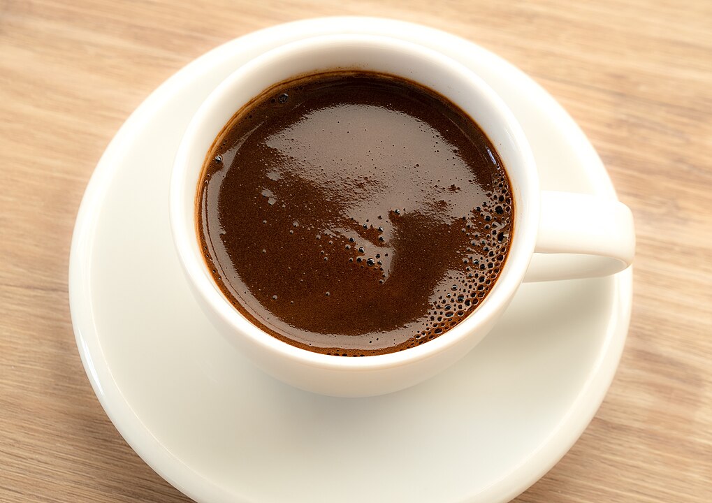
    However, she cautions that this remains a hypothesis, not a treatment protocol. Chocolate isn’t a cure-all, and overindulgence can lead to acne, migraines, or weight gain. But if jealousy creeps up with your morning coffee—a small piece of dark chocolate might just help.

  • The Mechanism of Fear: How to Train Your Brain Not to Be Afraid

    The Mechanism of Fear: How to Train Your Brain Not to Be Afraid

    Imagine you see a stray dog running toward you. At that moment, the image of the dog, the sound of its running, and other sensory information is transmitted through the thalamus and cerebral cortex to the amygdala—the brain’s emotional center.

    The amygdala is a paired structure deep within the brain, consisting of several nuclei. Two nuclei are responsible for fear: the lateral and central. The lateral nucleus functions as a receiver, taking in information from other structures. The central nucleus acts as a transmitter, sending commands about what to do next.

    Your amygdala decides that the running dog is dangerous and sends signals to other brain structures:

    • Hypothalamus. It causes the adrenal glands to release the hormones adrenaline and noradrenaline into the bloodstream, preparing your body for fight or flight: you begin to sweat, your pupils dilate, your breathing quickens, blood rushes to your brain and muscles, and digestion slows down.
    • Periaqueductal gray matter. This makes you freeze in place, like a deer in headlights. It might seem like a pointless reaction—you’d be better off looking for a rock or stick to fend off the dog. But your brain doesn’t think so. Millions of years of evolution tell it that freezing is an advantageous strategy. The predator might pass by, and you won’t have to waste energy fleeing, risking becoming someone’s lunch.
    • Paraventricular nucleus of the hypothalamus. This structure orders the secretion of cortisol—the stress hormone. It conserves energy to help you endure the dangerous situation. Additionally, cortisol allows the amygdala to work at full capacity: since the situation is dangerous, you need to react to any frightening stimuli, and the amygdala excels at this.

    Let’s say the dog really was dangerous, barked at you, or bit you. A strong connection between the image of the animal and the pain of the bite has now formed in your amygdala. From now on, the sight of a dog running toward you will trigger fear, even if it’s a friendly neighborhood dog. Moreover, each new episode of fear triggered by a dog will strengthen the neural connections in the amygdala and hippocampus, along with your fear of man’s four-legged friends.

    But this doesn’t mean you’ll panic at the sight of a dog for the rest of your life. Thanks to neuroplasticity—the brain’s ability to strengthen and weaken connections between neurons—you can overcome your fear.

    How to Overcome Fear

    Retrain Your Brain Through Action

    As mentioned above, the central nucleus of the amygdala actively participates in creating fear: it connects safe stimuli with presumably dangerous ones and sends signals to other brain structures. Because of this nucleus’s work, your neighbor’s dog, which has never bitten you, makes your heart beat faster and your palms sweat.

    In his book Taming the Amygdala, John Arden explains that the central nucleus can be overcome by another part of the amygdala—the bed nucleus of the stria terminalis. To activate it, you need to take concrete action, such as petting your neighbor’s dog.

    Furthermore, action activates the prefrontal cortex. Then the following happens: signals continue to arrive at the lateral nucleus of the amygdala, but the active prefrontal cortex suppresses the connection between the lateral and central nuclei. As a result, no commands come from the central nucleus—fear doesn’t arise.

    If you want to get rid of fear, confront it head-on.

    Want to overcome your fear of dogs? Get your own or play with a friend’s dog. The prefrontal cortex will assess the situation and prevent the amygdala from expressing fear. Eventually, the image of a dog will lose its “danger” label, and you’ll stop trembling at the sight of one.

    How long you’ll need to play with someone else’s dog and whether the fear will return if you suddenly see a stray dog depends on how long you’ve been afraid.

    Do It Quickly

    The sooner you take a step toward your fears, the better. Each episode of fear strengthens neural connections in the amygdala, making it increasingly difficult to overcome.

    The ideal time to fight fear is the first week after it forms. Scientists at McGill University discovered that fear extinction is related to CP-AMPAR receptors in the neurons of the lateral amygdala.

    In the first 24 hours after a new fear forms, the number of these receptors increases, then returns to normal levels within a week. After that, the fear becomes firmly established and becomes harder to fight.

    In experiments with mice, scientists identified the ideal scheme for fighting fear: within the first 24 hours after its formation, you need to see the frightening stimulus again, then work on unlearning the fear. For example, first you watch a video of an angry dog, then half an hour later you pet a friendly neighborhood dog.

    The video activates the fear and ensures neuronal plasticity, while playing with the dog helps eliminate the fear. However, this scheme only works in the first week, while the CP-AMPAR receptors haven’t returned to their previous levels. If you miss the window for working with the fear, it will be much harder to completely overcome it.

    To prevent fear from becoming entrenched, try to overcome it as quickly as possible.

    Activate the Prefrontal Cortex

    Since the prefrontal cortex can suppress excessive amygdala activity, activating it helps fight fear and anxiety.

    There are two proven ways to “turn on” this brain region:

    • Exercise. Physical activity increases prefrontal cortex activity.
    • Meditate. Meditation increases gray matter in the prefrontal cortex and decreases it in the amygdala. That’s why Buddhist monks are so calm: after many years of practice, their amygdala has shrunk and stopped being frightened by everything. However, a single meditation session won’t help: you’ll need to meditate for at least eight weeks, 40 minutes a day, for structural brain changes to occur.

    Remember: meditation and exercise will help you fight anxiety, but won’t rid you of already-formed fears. You can only do this by deliberately placing yourself in a similar stressful situation that ends favorably.

  • Can You Die From Fear?

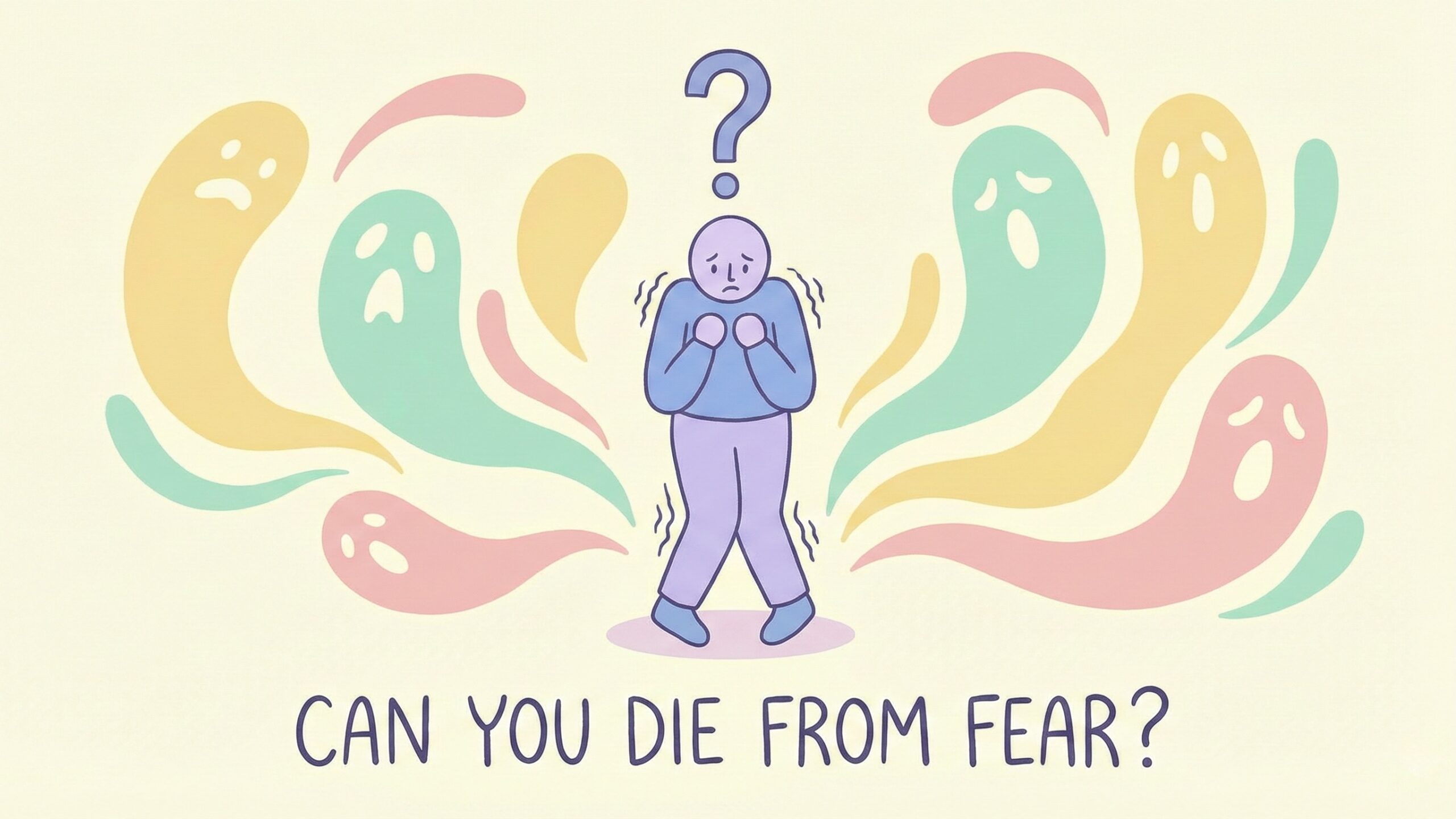
    Can You Die From Fear?

    We all sometimes say things like “you scared me to death!” But can intense fear actually lead to death?

    Humans, like all other animals, have an evolutionarily developed protective mechanism of “fight or flight” that our bodies activate automatically during intense stress. This was described by Dr. Walter Cannon, a professor in the Department of Physiology at Harvard University, in 1915.

    When we face danger, the autonomic (involuntary) nervous system responds. At its command, the adrenal glands release adrenaline, which rapidly spreads throughout the entire body.

    This hormone causes the opening of calcium channels in the membranes of heart muscle cells. Calcium ions make the heart contract faster. Blood flow to the muscles increases, pupils dilate, and the digestive process slows down. The body prepares for a fight or quick escape to protect itself.

    This helped our primitive ancestors better cope with dangers like a jaguar suddenly jumping out of the bushes.

    However, releases of adrenaline, despite their usefulness in moments of danger, lead to risky consequences. An overload of cells with this hormone can lead to a situation where the heart muscle simply cannot relax, which leads to arrhythmia and ventricular fibrillation. From truly intense terror, the heart can literally stop. Moreover, this happens even to completely healthy people.

    Not only fear but also other excessively strong emotions can lead to death. High levels of adrenaline can also kill during sexual intercourse, religious ecstasy, or excessive enthusiasm among sports fans. The number of heart attacks increases during tsunamis, hurricanes, earthquakes, and other natural disasters.

    And even if fear, stress, and anxiety don’t kill immediately, they contribute to the development of heart disease later on. For example, military conflicts become the cause of heart attacks and cardiac ischemia even years after the restoration of peaceful conditions.

    Japanese doctors in 1990 described the so-called takotsubo stress cardiomyopathy, when due to the strong impact of adrenaline and noradrenaline, myocardial contractility decreases. The disease is also called “broken heart syndrome,” as its trigger is intense emotional stress—the loss of a loved one, a job, means of subsistence, and so on.

    Perhaps the reason for the deadliness of fear is that modern people fight their terrors without resorting to a physical reaction.

    We stopped “fighting or fleeing” because stresses are now less material than predators or natural cataclysms.

    Surges of adrenaline, noradrenaline, and cortisol in our bodies don’t serve real needs—to become faster, stronger, more aggressive—but burden the body’s systems in vain.

    So fear can indeed literally lead to a heart attack. But fortunately, the probability that a person will simply up and die from terror is very, very small. To cope with this feeling in critical moments, simply breathe deeply. This helps normalize your pulse.

    And dedicate time to sports and aerobic exercises, because the cardiovascular system of trained people best handles surges of adrenaline.

  • Is Fear Really Contagious?

    Is Fear Really Contagious?

    Rapid heartbeat, sweaty palms, stomach cramps, trembling knees—the iron grip of fear is unmistakable. It’s one of the least controllable emotions, making it very difficult to suppress or hide. But can you catch fear if someone near you is frightened? The answer to this question lies in the nature of the phenomenon.

    What Function Does Fear Serve?

    The ancient mechanism of fear is activated mainly in the face of physical threats. If one person fears a predator, others notice and react, even if they don’t see the danger directly. By learning from others what to fear, people survive.

    In theory, fear should be contagious.

    When a person senses someone else’s fear, the amygdala activates—a brain region that plays a key role in forming emotions and responding to threats. A signal is then transmitted to the hippocampus to mobilize the nervous system. The body releases adrenaline, which speeds up the heartbeat and breathing. This is all part of the “fight or flight” response, which provides the strength and energy to confront threats or escape from them.

    How We Copy Fear

    People naturally mimic each other, and many emotions are inherently social. We not only experience them but also share them. Copying emotions, including fear, is a fundamental element of our mutual understanding.

    Research shows that we can feel fear simply by observing those who are afraid of something. Even if frightened faces flash before our eyes too quickly for us to consciously perceive them, the amygdala in our brain can still activate.

    This suggests that fear can spread subconsciously.

    In other words, research data indicates that we can copy fear from those around us, even if we haven’t been in the same dangerous situation or aren’t aware of what’s happening.

    Animals, such as certain rodents, also pick up fear from each other. When rats hear distress signals from other rats experiencing pain, they freeze in terror.

    Overall, fear can be transmitted through various pathways. Humans rely heavily on sight and hearing, while animals depend more on smell and hearing.

    Can We Smell Fear?

    Animals react acutely to odors. Many creatures—from zebrafish to ants—release pheromones when frightened or injured. These olfactory alarm signals warn of potential danger and trigger fear in other members of the same species.

    For years, scientists doubted that we could smell fear. After all, we don’t have an organ that detects pheromones. However, recent research provides grounds to suppose that humans can still sense the smell of fear through chemical signals produced by the brain that activate the amygdala.

    In a series of experiments, scientists collected sweat from those preparing to parachute jump and from those exercising on a treadmill. Participants were then given cotton pads with sweat samples to smell while undergoing brain scans. Test subjects who inhaled the sweat from parachutists showed increased amygdala activity. That is, they could likely pick up the parachutists’ emotions on a subconscious level.

    What Determines Our Reaction to Others’ Fear?

    All people share a common brain structure that makes fear contagious, but not everyone reacts to it the same way.

    Genetics, past experience, and level of empathy can influence this.

    People who aren’t particularly empathetic may experience less intense fear themselves when in the presence of someone gripped by terror. More empathetic individuals, conversely, will be affected more in such situations.

    When everyone around is panicking, those with relevant knowledge and experience are also likely to be less frightened. For example, if a wild snake escapes from a terrarium at the zoo, the crowd’s fear won’t affect an experienced zoologist who knows the reptile isn’t actually dangerous.

    Some people are biologically more inclined to pick up fear emotions from others. This may be connected to changes at the genetic level found in children whose parents survived catastrophes or wars. For instance, descendants of Holocaust victims possess heightened sensitivity to danger and therefore may not only experience fear more frequently themselves but also be more susceptible to others’ anxiety.

    So, fear is indeed contagious. First and foremost, this increases our chances of avoiding physical danger. But in everyday life, we also fear situations that don’t actually contain real threats, such as public speaking or job interviews, and other people can catch and pass on our panic moods further. Therefore, it’s worth remembering that fear is contagious.

  • Yes, Fish Can Drown, But How Does This Happen?

    To understand this, we first need to recall how fish breathe. No, fish don’t breathe water—they breathe air, just like you and me. We’re not so different from a hake after all! But when you’re at the bottom of the ocean, finding a breath of oxygen isn’t exactly easy. Fortunately, these vertebrates have an organ that allows them to extract dissolved oxygen from water: gills.

    Their functioning is as simple as it is remarkably efficient: water enters through the mouth, flows over the gill filaments where oxygen is absorbed while carbon dioxide is expelled. And the water? It exits through the gill openings as if nothing happened. An astonishing mechanism, adapted according to marine species… and one that sometimes fails to the point of drowning.

    More Suffocation Than Drowning

    Technically, there’s a catch. If we open the dictionary, the definition of “drowning” is “asphyxiation by the influx of liquid into the respiratory tract, particularly following immersion in water.” In theory, the fish is already immersed, and plunging it into water won’t cause drowning. However, there are several scenarios where fish can lack oxygen underwater. In these cases, we’re talking more about suffocation, or drowning in a non-traditional sense of the term.

    Generally speaking, if a fish’s gills are damaged, obstructed, or deprived of oxygenated water, suffocation occurs. Drowning is inevitable. And as you’ll see, this is far from an isolated phenomenon.

    Returning to those famous gills, as impressive as they are, they have a flaw for certain species: to maintain water flow over their gills, these species must swim continuously. This is the case, for example, with most sharks. If they become stuck or caught in a fishing net, immobilization deprives them of oxygen and they drown. And what about a plastic bag blocking a tuna’s mouth? Once again, drowning.

    Some species, like the Siamese fighting fish, have also developed a very particular organ called a “labyrinth,” which allows them to breathe atmospheric air. This is a way to survive in their oxygen-poor environment: marshes, rice paddies, puddles, or stagnant waters, also called hypoxic zones. A very handy trick… that can also lead to drowning! Strange as it may seem, if they don’t have access to the surface to breathe, they die by drowning.

    Too Many Nutrients, Less Oxygen in the Water

    There’s one phenomenon in particular that results in killing all aquatic life by “drowning”: eutrophication. Unfamiliar with the term? You’re far from alone. And yet, it should be well-known to humans, because they’re often the primary actors behind it.

    Eutrophication occurs when the concentration of phosphorus or nitrogen increases in water, which then accumulates far too many nutrients. This causes excessive and massive proliferation of algae. As these algae decompose, they consume enormous amounts of oxygen—all the available oxygen, in fact. The result: all fish that no longer have oxygen for their gills die of asphyxiation.

    What’s the connection to humans? It’s very often industrial and agricultural activities, as well as domestic uses, that cause this accumulation of nutrients in water and the resulting chain reaction. And when it’s not humans’ fault, it’s… hippos’.

    Inadvertently, these large mammals kill thousands of fish each year. Their excrement, released shamelessly into rivers, is decomposed by bacteria that once again consume enormous amounts of oxygen. In the end, it’s the fish that pay the price, deprived yet again of this vital gas.

  • Human Immunity Is Partly Inherited From Bacteria

    Human Immunity Is Partly Inherited From Bacteria

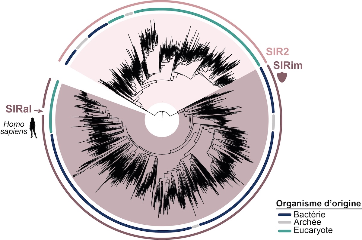
    The living world is rich in diversity, but all organisms share one thing in common: the evolutionary pressure exerted on them by pathogens. To defend themselves, bacteria, plants, animals, and others have developed a wide array of mechanisms over millions of years of evolution. To date, nearly 250 bacterial anti-infectious systems are known, which rely on immune proteins, some of which show surprising similarities to human immune proteins. By examining these connections, Enzo Poirier from Inserm and the Curie Institute in Paris, Aude Bernheim from the Pasteur Institute in Paris, and their colleagues discovered that numerous proteins involved in innate immunity in humans are derived from bacterial proteins.

    To defend against viruses, certain bacteria rely on the SIR2 (Silent Information Regulator 2) protein domain. A protein domain is a part of a protein that adopts a structure independently from the rest of the molecule. When a virus infects a bacterium, SIR2 disrupts the cell’s metabolism by destroying a molecule essential to it, thereby causing the cell’s death. SIR2 thus prevents viral replication and simultaneously protects the rest of the bacterial colony. Through extensive phylogenetic work—that is, the study of evolutionary relationships between organisms—the biologists identified a domain homologous to SIR2 in humans, called SIRa1. They then went further and demonstrated in mouse and human immune cell models that this domain plays a crucial role in innate immunity, the first line of defense against viral or bacterial infection. Its function? It serves as a pivot in the TLR (Toll-like receptor) signaling pathway, which enables the detection of pathogens during infection and triggers an inflammatory response. By reducing or eliminating SIRa1 production in mice, Enzo Poirier, Aude Bernheim, and their colleagues observed that the inflammatory response to viral or bacterial infection was severely impaired and dysfunctional.

    Interestingly, the SIRa1 domain appears not in just one protein, but in an entire ancient and diverse protein family whose members the team found in no less than 19% of the eukaryotes—organisms composed of cells with nuclei, such as plants or animals—that they analyzed. This means that the associated immune mechanisms, derived from bacteria, have been very widely conserved throughout evolution and across the living kingdom, since bacteria and eukaryotes constitute two of its three major groups. And that the underlying molecules have retained their structure and function over billions of years!

    This finding establishes the framework for the emerging field of ancestral immunity, which aims to systematically identify evolutionary links between immune molecules in bacteria and eukaryotes that share a distant common origin. Characterizing these proteins and better understanding them could help outline new therapeutic strategies, particularly against certain autoimmune diseases that result from overactivation of TLR receptors.

  • What Coffee Really Does to Our Brains at Night

    What Coffee Really Does to Our Brains at Night

    The brain follows a precise sequence of phases to repair, sort, and reinforce what the day has imprinted. In this fragile balance, even a familiar molecule like caffeine is enough to shift everything out of place. Consumed a few hours before bedtime, it interferes with the electrical signals meant to calm mental activity and interrupts the deep rhythms essential for rest. On the surface, sleep may seem to occur normally, but the invisible alterations become measurable at the very core of the brain. It is at this uneasy crossroads between coffee and sleep that researchers are now focusing their attention.

    Caffeine Blocks the Brain from Entering Deep Rest

    During sleep, the brain cycles through different stages, some of which are essential for physical and mental recovery. One of the most important is slow-wave sleep, marked by highly structured brain activity dominated by slow oscillations. This is when the brain reorganizes, erases unnecessary overload, and stabilizes recent learning.

    A dose of caffeine equivalent to two cups of coffee disrupts this natural process, as observed by researchers at the University of Montreal. By recording brain electrical signals in forty volunteers, they found that the brain maintains heightened activity, incompatible with fully restorative sleep. The results showed a clear reduction in delta and alpha waves, which usually signal deep cerebral relaxation.

    The authors of the study, published in Communications Biology, describe a shift into a state of “criticality.” This state lies at the boundary between order and chaos. While useful during the day for decision-making and processing new information, it becomes problematic at night, preventing the brain from slowing down. The body enters a kind of silent wakefulness—imperceptible to the sleeper but very real in neural connections.

    When Coffee and Sleep Clash in Young Adults

    The effects of caffeine on sleep are not uniform. The study found that young adults, aged 20 to 27, are significantly more sensitive to nighttime caffeine stimulation than older individuals. In their case, brain signals measured during the night were more disrupted, particularly during REM sleep. This phase, in which the brain generates vivid mental experiences such as dreams, also plays an important role in emotional balance and creativity.

    This sensitivity is partly explained by the higher number of adenosine receptors in younger brains. Adenosine is a naturally produced molecule that signals fatigue and acts as a neuronal brake. Caffeine occupies these receptors without activating them, blocking the fatigue signal and artificially maintaining wakefulness. According to researchers cited by Science Alert, this stronger interaction in younger brains makes their sleep more vulnerable—even when falling asleep doesn’t seem to be affected on the surface.

    Students and young professionals, who often consume coffee to stay alert in the evening, may be particularly at risk. The reality is that even if they manage to fall asleep after drinking coffee, the quality of their sleep is deeply compromised, without them realizing it.

    Brain Waves Altered Even During Memory Phases

    The brain never fully sleeps. Throughout the night, it sorts memories, strengthens certain circuits, weakens others, and stabilizes what was learned during the day. This sorting and strengthening relies on slow, well-orchestrated electrical rhythms, mostly seen during deep sleep. These rhythms allow neurons to synchronize, transferring information from the cortex to the hippocampus, the brain region crucial for memory.

    When the brain is under the influence of caffeine, this harmony breaks down. The study indicates that theta and delta waves—central to memory consolidation—become less frequent and less intense. In their place, faster waves appear, associated with wakefulness or mental agitation. This disorganization undermines the brain’s ability to fix memories or process recent learning.

    Even without insomnia, short-term memory quality can deteriorate after a caffeine-influenced night. These alterations are not immediately visible, but their repetition over time could weaken certain cognitive functions, particularly in those who regularly drink coffee late in the day.

    Sleep Altered in Depth Without Awareness

    What makes caffeine unique is that it disrupts sleep subtly. It doesn’t necessarily cause nighttime awakenings or outright insomnia. Its deceptiveness lies in making us believe we’ve slept well, while brain activity remains disorganized at a deeper level.

    According to data from the researchers, caffeine also changes the way electrical signals fluctuate throughout the night. The brain loses part of its usual stability, showing a global increase in entropy—that is, greater complexity and unpredictability in signals. This rise is useful when awake, reflecting high intellectual responsiveness. But during sleep, it represents a form of overactivity that prevents the brain from regenerating properly.

    This phenomenon was analyzed using machine learning techniques by the study’s authors. The data revealed that complexity markers—such as spectral entropy or signal compressibility—were the most sensitive to caffeine ingestion, even more than simple wave variations. This result highlights just how insidious the effect is. It doesn’t change the sleeper’s perception but profoundly alters how the brain recovers.

  • Why Morning Coffee Really Does Make You Happier, According to Science

    Why Morning Coffee Really Does Make You Happier, According to Science

    It only takes an aroma to evoke the warmth of an ordinary morning, the promise of a new beginning. Coffee has established itself as the faithful companion of waking up, far beyond being just a stimulant. If this daily gesture brings so much comfort, it is not only a matter of taste or routine. The effects of coffee on mood are beginning to reveal their secrets, and science is paying close attention.

    Coffee: a universal morning ritual

    Every morning, billions of people unknowingly share the same gesture. If this ritual is so deeply rooted, it is not just about taste or habit. It is also a matter of the brain.

    A study conducted by the Universities of Bielefeld and Warwick on more than 230 young adults revealed that positive emotions—such as enthusiasm or a sense of well-being—were significantly enhanced after drinking coffee in the morning. These results did not come from a closed laboratory. Participants were followed in their daily lives through an app that asked them to rate their mood seven times a day, specifying whether they had consumed a caffeinated beverage in the previous 90 minutes. This protocol, known as the “experience sampling method,” allows for greater ecological validity.

    The researchers observed that the link between caffeine and mood was particularly strong within the first two and a half hours after waking. Beyond that window, the positive effects faded, only to reappear slightly at the end of the day. This consistency sheds new light on the biological dimension of this small morning pleasure.

    Coffee’s effect on mood depends on timing

    Why does caffeine act more strongly in the morning? Part of the explanation lies in our chronobiology. During the night, our level of adenosine increases. This molecule slows neuronal activity and promotes sleep. By blocking adenosine receptors, caffeine triggers greater release of dopamine and norepinephrine, which increases alertness and supports a more positive mood. The effect is all the more pronounced because the body is emerging from a long caffeine fast, as is often the case upon waking.

    It is also possible that morning coffee has a stronger psychological impact. Simply anticipating this comforting moment could trigger an enhanced placebo effect, amplifying caffeine’s chemical action. Moreover, according to the study published in Scientific Reports, this well-being peak does not depend on habitual consumption levels, sleep quality, or even pre-existing anxiety or depressive states. It is an almost universal response.

    The study’s authors also note that coffee seems to enhance positive emotions more than it reduces negative ones. For example, it increases feelings of contentment but does not significantly affect anxiety. This suggests that caffeine acts as an amplifier of pleasant emotions, rather than as an anxiolytic.

    A brain mechanism that goes beyond taste

    What this research confirms is that the effects of coffee on mood are not solely due to conditioning or reverse withdrawal. Even people who are not very dependent on caffeine experience a positive impact, which contradicts the theory that only the relief of withdrawal symptoms is at play.

    Another factor comes into play: the social context. Interestingly, the emotional benefits of coffee were less intense when participants were surrounded by other people. This may be because social stimulation already acts as an emotional activation lever, reducing the margin of coffee’s impact. Conversely, when subjects reported being more tired than usual, caffeine triggered a clearer improvement in their mood.

    The Bielefeld researchers nevertheless emphasize caffeine’s limits. While it temporarily improves mood and alertness, it can disrupt sleep if consumed late in the day. And excessive consumption is still associated with known risks, such as irritability or dependence. That said, these side effects appeared only in a minority of individuals in the findings relayed by SciTechDaily, and did not undermine the positive link between coffee and morning mood.

    Thus, science confirms what instinct long suggested: coffee, taken at the right moment, is more than just a drink. It acts as a catalyst for energy and good humor—a true biological and emotional lever for starting the day right.

  • What Causes Brain Freeze?

    What Causes Brain Freeze?

    Have you ever felt a sharp pain in the frontal lobe of your brain, like a cold stabbing sensation, while enjoying delicious ice cream on a hot summer day or drinking cold water? If you’re reading this article, you probably have.

    buy cialis super active online https://cphia2023.com/wp-content/uploads/2025/03/jpg/cialis-super-active.html no prescription pharmacy

    If so, don’t worry – you don’t need to see a doctor unless you experience this pain frequently or for extended periods. But don’t be concerned if you haven’t felt it either.

    According to research, approximately 40% of the population experiences this pain, with it being more common among those who suffer from migraines; meaning the majority never experience this pain. Children are more prone to experiencing brain freeze as they may consume cold foods and drinks too quickly. The medical term for this sensation is Sphenopalatine ganglioneuralgia, but as you might have noticed, “brain freeze” is a much simpler name.

    The Cause of Brain Freeze

    Brain freeze typically occurs when you consume things like ice cream, very cold drinks, ice cubes, or when you inhale freezing cold air. According to research conducted in 2012 on the cause of brain freeze, this phenomenon is entirely related to blood flow. When something cold is consumed rapidly, if it suddenly and continuously contacts the palate, it triggers the trigeminal nerve (which connects the brain and face) by constricting the blood vessels leading to the brain.

    buy topamax online https://silvermancare.com/wp-content/uploads/2025/03/jpg/topamax.html no prescription pharmacy

    The brain, disturbed by the sudden temperature change, directs warm blood to these constricted vessels. The rush of blood to these cold-constricted vessels is ultimately perceived by the nerves as pain caused by cold – that is, “brain freeze.”

    According to research, the width of the area touched by the cold, the number of points contacted simultaneously, and the speed of entry are more important for brain freeze than the temperature itself. This is why ice cream of the same temperature is more likely to cause brain freeze when eaten quickly or spread throughout the mouth. The same applies to iced water.

    The Solution to Brain Freeze

    The way to get rid of brain freeze is quite simple. All you need to do is eliminate the cause – the cold. Since brain freeze is a “referred pain,” meaning that data input at one point causes a pain signal at another point, changing the inputs directly affects the pain. Solutions for brain freeze include stopping the consumption of the cold food or drink, moving away from the cold environment, drinking warm water, or pressing your thumb against your palate for heat transfer. Taking aspirin or painkillers won’t be very effective in alleviating brain freeze because in 98% of people, this pain lasts less than 5 minutes. By the time the medication takes effect, the brain freeze will have already passed.

    Brain Freeze and Other Headaches

    Unlike some other headaches that cause stomach pain or sensitivity to light/noise, brain freeze has no effect other than a stabbing sensation in the brain. If you experience vision loss, dizziness, difficulty moving or speaking along with brain freeze, it is recommended to seek help immediately. Additionally, it was found that those who suffer from various headaches and/or children whose parents have experienced brain freeze are more prone to experiencing brain freeze. Other types of headaches experienced by parents were not related to the child experiencing brain freeze, but children of parents who never experience headaches were also less likely to experience brain freeze.

    buy keflex online https://cphia2023.com/wp-content/uploads/2025/03/jpg/keflex.html no prescription pharmacy

    So, even though it might seem funny, brain freeze could help us solve the heritability and causes of other types of headaches, such as migraines.
    buy lopressor online https://cphia2023.com/wp-content/uploads/2025/03/jpg/lopressor.html no prescription pharmacy

    Additionally, brain freeze may not only be related to humans. For example, the reactions of cats after being fed ice cream (although not recommended) and their neuroanatomy similar to ours may indicate that cats can also experience brain freeze.

    buy robaxin online https://silvermancare.com/wp-content/uploads/2025/03/jpg/robaxin.html no prescription pharmacy

  • The World as Seen by Europeans Before 1492

    The World as Seen by Europeans Before 1492

    On the eve of what historiography has termed the “Age of Discovery,” the Christian West held a geographical worldview that blended Greek, religious, and empirical influences, tinged with a sense of mystery. This perspective was far removed from the transformative understanding of the world that would emerge from the explorations and conquests of the 16th and 17th centuries, which decisively altered how the world was perceived and ushered in modernity. The first explorers, such as Christopher Columbus, were still men of the Middle Ages.

    “T-O” Mappa Mundi

    The Hereford Mappa Mundi, about 1300, Hereford Cathedral, England
    The Hereford Mappa Mundi, about 1300, Hereford Cathedral, England. Credit: Public Domain

    The “T-O” mappa mundi model is the primary representation of religious influence on medieval geography. Drawing inspiration from ancient scholars and biblical narratives, Church Fathers like Isidore of Seville (560-636) divided the world among the three sons of Noah: Asia to Shem, Europe to Japheth, and Africa to Ham.

    buy rybelsus online https://cialisnextdaydeliveryusa.com/buy-rybelsus.html no prescription pharmacy

    This geographical vision depicted the Earth as flat, contributing to the misconception that medieval people were unaware of the planet’s roundness (despite this knowledge dating back to antiquity and being studied by medieval scholars familiar with Greek science). The East was placed at the top, symbolizing Eden, the Paradise.

    The “T-O” model persisted until the end of the Middle Ages, with the three sons of Noah occasionally being replaced by the Three Magi, each symbolizing a “continent,” even though the term “continent” itself is more characteristic of the modern era.

    The Role of Portolan Charts

    Marine charts, which developed significantly from the 13th century onward, were crucial in understanding how Europeans viewed the world at the time. Originating from Italian thalassocracies (Pisa, Genoa, Venice), portolan charts were used for Mediterranean navigation as early as the 13th century. These charts, often drawn on parchment, featured coastlines, ports, and hazards, and incorporated the use of the compass.

    buy colchicine online https://cialisnextdaydeliveryusa.com/buy-colchicine.html no prescription pharmacy

    A network of lines drawn on the sea allowed navigators to reach ports marked on the chart.

    Initially created for practical navigation and based on precise observations, portolan charts quickly became collector’s items, often idealized representations that blended cartographic details with figurative iconography, displayed in studiolos and princely palaces. They were richly decorated with geometric and floral motifs, coats of arms, ships, animals, and other designs.

    buy synthroid online https://cialisnextdaydeliveryusa.com/buy-synthroid.html no prescription pharmacy

    These charts essentially served as catalogs of ports, with names labeled in red or black according to their importance, but without indicating distances between them. Major rivers and sometimes topographical features were schematically represented, without accurate proportions.

    The World Map Before 1492

    The influence of Greek thought was central to the geographical understanding of the world. By the 15th century, the vision of Ptolemy, a 2nd-century Greek geographer, had become the most popular, gradually supplanting the flat, religious worldview of earlier centuries with the concept of a spherical Earth, as proposed by Eratosthenes. According to Ptolemy, the Indian Ocean was enclosed, a feature reflected in 15th-century maps, such as that of Nicolaus Germanus (1482).

    In 1450, the monk Fra Mauro created a mappa mundi that synthesized various cartographic traditions, both Christian and Arab, incorporating empirical information from early explorations of the time. While influenced by Ptolemy, Fra Mauro rejected the idea of an enclosed Indian Ocean.

    However, the most striking example of the 15th-century worldview, bridging the Middle Ages and the modern era, is undoubtedly the Behaim Globe. Inspired by Ptolemy, it integrated information from the travels of Marco Polo and John Mandeville, whose accounts would later guide Christopher Columbus himself.

    The Behaim Globe depicted a narrowed Atlantic Ocean, which, if crossed, would lead directly to Cipango (Japan) and Asia. In the middle, it was missing just the New World, which would be “discovered” a few months later by the Genoese navigator…

  • Science Sleep: Men and Women Do Not Sleep the Same Way

    Science Sleep: Men and Women Do Not Sleep the Same Way

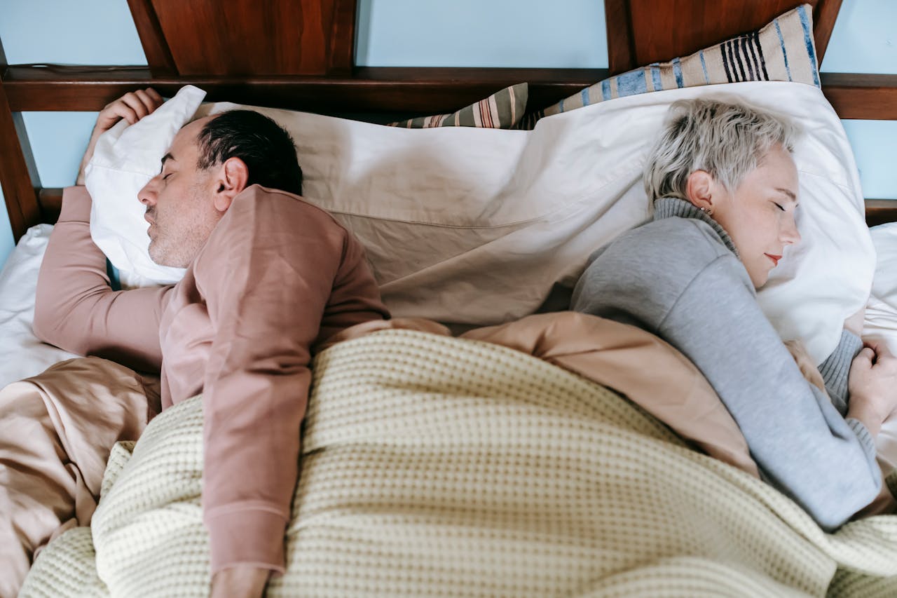
    Moms know it well: no one can sleep as deeply as a dad when a newborn cries at night. Now, a scientific study conducted on mice confirms that it’s not just dads who sleep better than moms, but men in general compared to women. Women, according to research published in Scientific Reports, tend to sleep fewer hours, wake up more frequently, and enjoy less restorative sleep compared to men.

    The Study:
    The researchers analyzed the sleep patterns of 267 mice. Male mice slept approximately 670 minutes (more than 11 hours) in a 24-hour period—an hour more than female mice.

    buy abilify online https://bereniceelectrolysis.com/jquery/js/abilify.html no prescription pharmacy

    This extra hour of sleep might correspond to the non-REM (nREM) phase, during which the body “repairs” itself. In addition to being shorter and less deep, the females’ sleep was also more fragmented compared to the males, with shorter rest periods and frequent awakenings.
    buy xenical online https://viagra4pleasurerx.com/buy-xenical.html no prescription pharmacy

    Survival of the species?
    What explains these differences between the sexes? According to Rachel Rowe, one of the study’s authors, biological factors play a crucial role. “Females are programmed to be more sensitive to their environment and to respond when necessary because they are typically the ones caring for the young,” explains Rowe, who jokingly adds, “If we women slept like men, wouldn’t our species go extinct?

    buy erythromycin online https://viagra4pleasurerx.com/buy-erythromycin.html no prescription pharmacy

    ”

    Men and women (aren’t) the same:
    From a scientific perspective, it’s essential that the female sex be adequately represented in sleep studies—something that hasn’t been done until now. While we know that sleep deprivation can have highly negative effects on health, are these effects the same for men and women if their sleep is already different to begin with?

    buy cialis black online https://bereniceelectrolysis.com/jquery/js/cialis-black.html no prescription pharmacy

    Perhaps, Rowe concludes, women stress unnecessarily about not sleeping enough, simply because they compare themselves to their male partners.
    buy provigil online https://viagra4pleasurerx.com/buy-provigil.html no prescription pharmacy

  • 10 Drugs That Changed the World

    10 Drugs That Changed the World

    Throughout the history of medicine, important pharmaceutical discoveries have changed the course of healthcare and the human experience. So chemotherapy drugs, penicillin, insulin, ether, chlorpromazine, thalidomide, contraception, idoxuridine, and azidothymidine are not just chemical molecules. They are symbols of the revolutionary forces that changed medicine and society. The history of these drugs is a story of technological progress, moral dilemmas, and social transformation, beginning with the development of anesthesia and ending with the eradication of infectious diseases.

    Ether

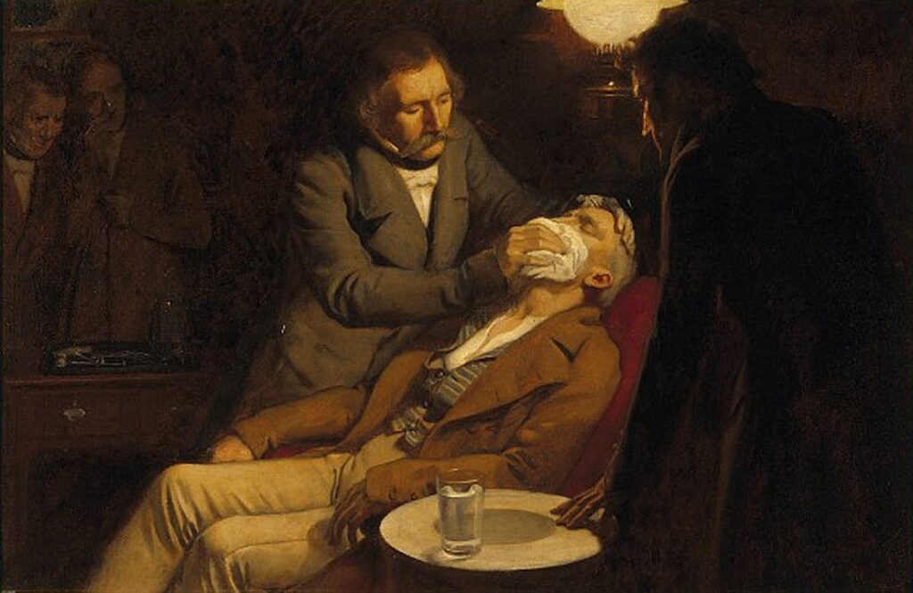
    V0018140 The first use of ether in dental surgery, 1846. Oil painting.Credit: Wellcome Library, London. Wellcome Images.images@wellcome.ac.uk.http://images.wellcome.ac.uk.The first use of ether in dental surgery, 1846. Oil painting by Ernest Board
    V0018140 The first use of ether in dental surgery, 1846. Oil painting.Credit: Wellcome Library, London

    Ether is a volatile liquid that can produce anesthesia when inhaled. Ether was first discovered in the 13th century by a Spanish chemist named Raymundus Lullus (Ramon Llull). However, its use as an anesthetic began in the mid-19th century. Crawford Long, a physician from Georgia in the USA, used it as a surgical anesthetic for the first time in 1842. He removed a tumor from the neck of a patient who was under the influence of ether. However, he did not publish his results until 1848.

    The first public demonstration of ether anesthesia was done by William Morton, a dentist from Boston, USA, in 1846 at the Massachusetts General Hospital.

    Aspirin

    Aspirin
    Bayer’s aspirin commercial.

    Aspirin is a drug that can relieve pain, inflammation, and fever. It is derived from salicylic acid, a compound found in the bark of willow trees and other plants. The use of salicylic acid as a medicine dates back to ancient times when people chewed willow bark or drank tea made from it to treat various ailments. In 1828, Johann Buchner, a German chemist, isolated salicin, a precursor of salicylic acid, from willow bark.

    In 1897, Felix Hoffmann, a German chemist working for Bayer, synthesized acetylsalicylic acid, a more stable and less irritating form of salicylic acid, and named it aspirin. Bayer patented the drug in 1899 and marketed it as a painkiller.

    Insulin

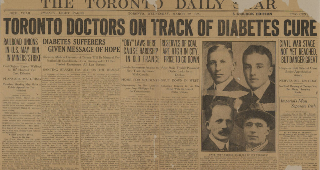
    Front page of the Toronto Daily Star, March 1922.
    Front page of the Toronto Daily Star, March 1922.

    Insulin is a hormone that regulates blood sugar levels. The pancreas, a gland located in the abdomen, is responsible for producing this hormone. In individuals with diabetes, insulin is either insufficient or ineffective, leading to elevated blood sugar levels and various complications. In 1889, German physiologists Oskar Minkowski and Joseph von Mering discovered that removing the pancreas from dogs caused them to develop diabetes. This suggested that the pancreas contained a substance that prevented diabetes.

    In 1921, three Canadian researchers, Frederick Banting, Charles Best, and James Collip, working at the University of Toronto, isolated insulin from the pancreas of dogs. They successfully lowered blood sugar levels by injecting insulin into diabetic dogs. In 1922, they achieved a successful treatment by administering insulin injections to a 14-year-old child with type 1 diabetes.

    Insulin became the first effective treatment for diabetes, saving millions of lives. Banting and the head of the Toronto laboratory, John Macleod, shared the Nobel Prize in Physiology or Medicine in 1923 for the discovery of insulin.

    Penicillin

    Penicillin
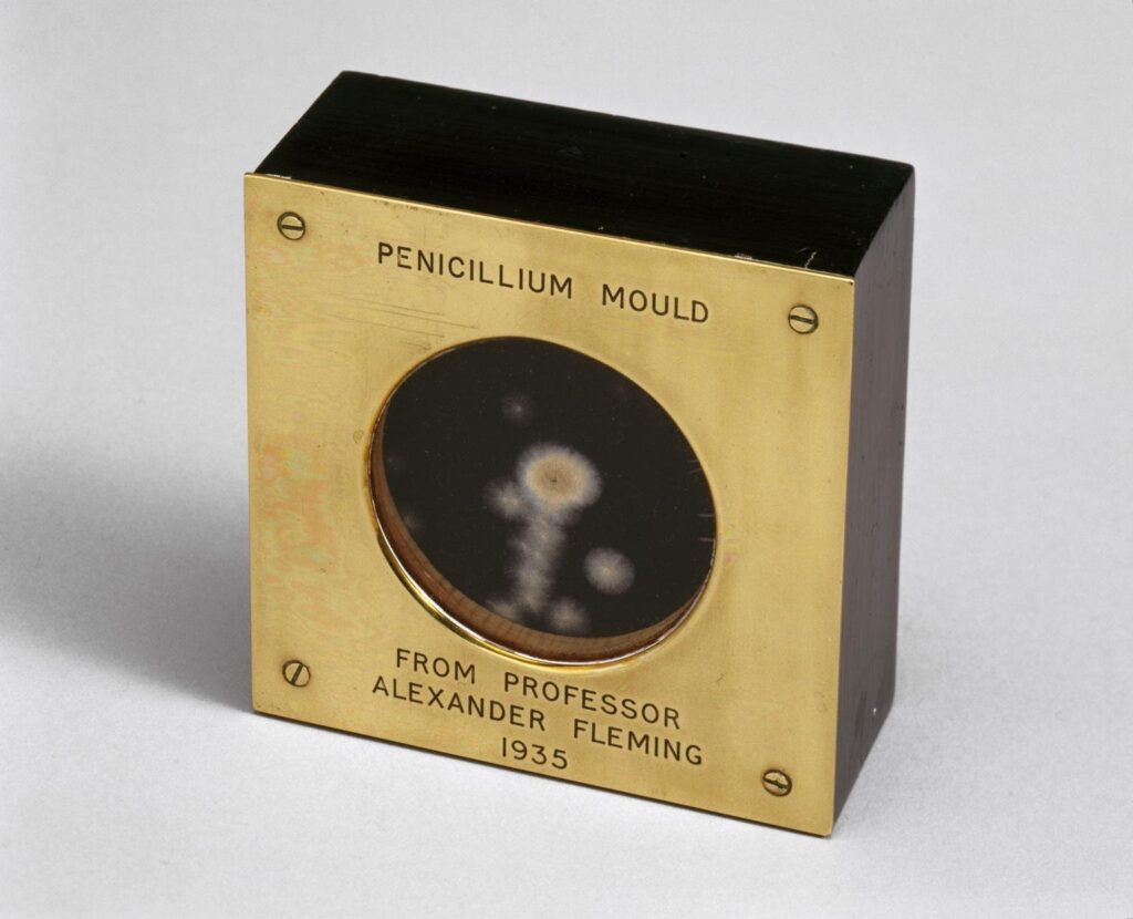
    A sample of penicillium mould, gifted by Alexander Fleming to a colleague at St Marys Hospital, London, 1935. Image: Science Museum.

    Penicillin is an antibiotic that can kill or inhibit the growth of certain bacteria. Penicillium is a type of mold that produces it. The discovery of penicillin is attributed to the Scottish bacteriologist Alexander Fleming, who worked at St. Mary’s Hospital in London.

    In 1928, Fleming discovered that mold had contaminated a Staphylococcus bacterial culture he had left on his workbench, preventing the bacteria around the mold from growing. He identified the mold as Penicillium and named the antibacterial substance it produced penicillin. However, he struggled to purify and mass-produce penicillin, and his discovery went largely unnoticed.

    In 1939, two biochemists from the University of Oxford, Howard Florey and Ernst Chain, revived Fleming’s work and developed a method to obtain and purify penicillin from mold cultures. They tested penicillin on mice and humans, demonstrating its effectiveness against various bacterial infections.

    During World War II, with the assistance of the U.S. and British governments and pharmaceutical companies, penicillin entered mass production and became a lifesaving drug for wounded soldiers and civilians. Fleming, Florey, and Chain shared the Nobel Prize in Physiology or Medicine in 1945 for the discovery and development of penicillin.

    Chemotherapy Drugs

    Chemotherapy drugs are medications that can kill or halt the growth of cancer cells, which are abnormal cells that divide uncontrollably and invade other tissues. The first chemotherapy drug, nitrogen mustard, is a chemical warfare agent that damages DNA and inhibits cell division.

    In 1942, pharmacologists Alfred Gilman and Louis Goodman from Yale University tested nitrogen mustard on mice with lymphoma, a cancer type affecting lymph nodes. They found that the drug reduced tumor size and prolonged the survival of the mice.

    In 1943, they treated a non-Hodgkin’s lymphoma patient with nitrogen mustard, achieving temporary remission. Nitrogen mustard became the prototype of alkylating agents, a class of chemotherapy drugs that interfere with DNA synthesis and function. Dr. Sidney Farber’s work in the 1940s laid the foundation for modern cancer chemotherapy.

    Later, new types of chemotherapy drugs were found or created. These include antimetabolites, which copy and damage the building blocks of DNA and RNA, vincristine and paclitaxel, which come from plants and stop cells from dividing, and cisplatin and carboplatin, which are made of platinum and stop DNA from replicating by creating cross-links in it.

    Chemotherapy drugs are often used in combination to enhance efficacy and reduce side effects.

    Chlorpromazine

    Chlorpromazine is a drug that can reduce the symptoms of psychosis, such as hallucinations, delusions, and agitation. It belongs to a class of drugs called antipsychotics or neuroleptics, which affect the activity of certain brain chemicals called neurotransmitters. Paul Charpentier, a chemist at the French pharmaceutical company Rhône-Poulenc, created chlorpromazine for the first time in 1950. It was originally designed as an antihistamine, a drug that blocks the effects of histamine, a substance that causes allergic reactions.

    In 1951, Henri Laborit, a French surgeon, tested chlorpromazine on surgical patients and found that it induced a state of calmness and indifference without causing a loss of consciousness. He suggested that the drug could be used to treat psychiatric disorders. In 1952, Jean Delay and Pierre Deniker, two psychiatrists at the Sainte-Anne Hospital in Paris, tried chlorpromazine on patients with schizophrenia, a severe mental disorder characterized by psychosis.

    They observed that the drug reduced the intensity and frequency of psychotic symptoms, such as delusions, hallucinations, and disorganized speech and behavior. Chlorpromazine became the first drug to treat schizophrenia and other psychotic disorders and opened a new era of psychopharmacology. Chlorpromazine also inspired the development of other antipsychotic drugs, such as haloperidol, clozapine, and risperidone.

    Thalidomide

    Thalidomide is a drug that can treat certain inflammatory and autoimmune diseases, such as leprosy and multiple myeloma, a type of blood cancer. It also has sedative and anti-nausea effects. However, taking thalidomide while pregnant is well known to lead to severe birth defects. Wilhelm Kunz, a chemist at the German pharmaceutical company Chemie Grünenthal, created the first synthetic version of thalidomide in 1953. It was marketed as a safe and effective sedative and antiemetic, especially for pregnant women suffering from morning sickness.

    However, in the late 1950s and early 1960s, thousands of babies were born with severe malformations, such as missing or shortened limbs, due to their mothers’ exposure to thalidomide during pregnancy. The drug was withdrawn from the market in 1961 after the link between thalidomide and birth defects was established by several researchers, including Frances Oldham Kelsey, a pharmacologist at the US Food and Drug Administration, who refused to approve thalidomide for sale in the US.

    Thalidomide became one of the biggest medical disasters in history and led to stricter regulations and ethical standards for drug testing and approval. In the 1990s, thalidomide was rediscovered as a treatment for leprosy and multiple myeloma after researchers found that it had immunomodulatory and anti-angiogenic properties, meaning that it could modulate the immune system and inhibit the formation of new blood vessels.

    Thalidomide was approved for these indications, under strict conditions and monitoring, in several countries, including the US. Thalidomide also served as a model for the development of other drugs with similar mechanisms of action, such as lenalidomide and pomalidomide.

    Birth Control

    Birth control is a term that refers to any method or device that can prevent pregnancy. Hormonal methods include birth control pills, patches, injections, implants, or intrauterine devices (IUDs), such as hormonal IUDs. Barrier methods include condoms, diaphragms, or cervical caps. Natural methods involve techniques like abstinence, withdrawal, or fertility awareness. Surgical options include vasectomy or tubal ligation.

    The history of birth control dates back to ancient times, when people used various herbs, plants, animal products, or physical methods to prevent pregnancy or induce abortion. Among the earliest documented forms of birth control are the use of vaginal suppositories by the ancient Egyptians, the use of the plant silphium by the ancient Greeks and Romans due to its contraceptive and abortifacient properties, and the practice of coitus interruptus or withdrawal, in various cultures.

    The development of modern birth control pills began in the 1950s, with key contributions from scientists such as Carl Djerassi, Gregory Pincus, and John Rock. The first oral contraceptive, Enovid, was approved for contraceptive use in 1960.

    Idoxuridine

    Idoxuridine is a drug that can treat herpes simplex keratitis, an eye infection caused by a virus. It is a nucleoside analogue, a modified form of deoxyuridine, that can block the replication of viral DNA. It was first synthesized by William Prusoff, a chemist at Yale University, in 1958. He initially developed it as an anticancer drug but later found that it had antiviral activity against the herpes simplex virus. In 1962, the US Food and Drug Administration approved it as the first antiviral agent.

    Azidothymidine

    Azidotimidin, also known as zidovudine or AZT, is a drug that can prevent and treat HIV/AIDS. It is also a nucleoside analogue, a modified form of thymidine, that can inhibit the enzyme reverse transcriptase that HIV uses to make DNA. Jerome Horwitz, a chemist at Wayne State University, created the first version of it in 1964. He intended it as a cancer therapy, but it proved ineffective and was shelved. In the 1980s, it was included in a screening program by the National Cancer Institute to identify drugs to treat HIV/AIDS. It showed promising results in laboratory and clinical trials, and it became the first drug to gain approval from the US Food and Drug Administration for treating AIDS in 1987.

  • 5 Popular Myths About Music

    5 Popular Myths About Music

    Music is so deeply woven into human culture and daily cognitive experience that folk wisdom, marketing campaigns, and misread scientific papers have generated enduring misconceptions about how sound affects the brain, animals, and physical matter. From parenting trends aimed at boosting infant intelligence to acoustic urban legends, examining five of the most widespread myths about music reveals the fascinating boundary between auditory science and popular belief.

    The Mozart Effect Makes Babies Smarter

    Few scientific misunderstandings have launched as many commercial products as the claim that playing classical music by Wolfgang Amadeus Mozart to infants or pregnant mothers permanently increases a child’s general intelligence. This phenomenon began in 1993 when researcher Frances Rauscher and her colleagues published a brief paper in the journal Nature. In their experiment, thirty six college students listened to ten minutes of Mozart’s Sonata for Two Pianos in D Major before taking a spatial reasoning test involving paper folding and cutting. The students scored temporarily higher than when they sat in silence or listened to relaxation tapes, an improvement that faded completely after fifteen minutes.

    Despite the researchers explicitly noting that the study only measured short term spatial arousal in adults, media outlets and toy manufacturers sensationlized the findings, culminating in the Governor of Georgia budgeting state funds in 1998 to hand out classical compact discs to every newborn in the state. Subsequent meta analyses involving thousands of participants demonstrated that there is no permanent increase in intelligence quotient from passive listening. Instead, the temporary cognitive boost stems from mood arousal: listening to any engaging audio, whether it is upbeat classical music, rock, or an interesting audiobook by Stephen King, wakes up the brain and briefly sharpens focus before a task.

    432 Hertz Is the Natural Healing Frequency of the Universe

    Across wellness forums and video platforms, countless tracks advertise music tuned to a reference pitch of A432 hertz, claiming that the modern international standard of A440 hertz is an unnatural tuning imposed in the twentieth century to induce anxiety and listener aggression. Proponents of the 432 hertz myth assert that this lower pitch aligns mathematically with the Schumann resonance of the Earth’s electromagnetic field, the geometry of ancient Tibetan singing bowls, and the golden ratio of the cosmos.

    Historical musicology and physics tell a much more practical story. Before the twentieth century, pitch standards across Europe varied wildly from town to town and century to century, depending on the physical size of local church organs and the temperature of concert halls. During the Baroque and Classical eras, tuning forks used by composers ranged anywhere from 409 hertz to well over 450 hertz. In 1859, the French government legally standardized pitch at 435 hertz to protect opera singers from straining their voices as orchestras tuned higher and higher for brighter acoustic projection, and in 1939 an international conference settled on 440 hertz as a convenient compromise for global radio broadcasting. Furthermore, the Schumann resonance actually fluctuates around 7.83 hertz rather than 8 hertz, and a hertz is simply a measurement of cycles per second based on an arbitrary human division of the solar day. While tuning an instrument down to 432 hertz creates a slightly warmer, mellower timbre due to reduced string tension, it holds no mystical biological superiority over 440 hertz.

    The Infamous 27 Club Is a Statistical Anomaly

    When iconic musicians such as Robert Johnson, Brian Jones, Jimi Hendrix, Janis Joplin, Jim Morrison, Kurt Cobain, and Amy Winehouse all passed away at the exact age of twenty seven, music journalism cemented the legend of the 27 Club, suggesting that rock and blues stars face a mysterious peak of mortality in their twenty seventh year. For decades, fans treated the number twenty seven as an ominous milestone for young artists living high risk lifestyles.

    In 2011, biostatisticians led by Adrian Barnett at the Queensland University of Technology tested this cultural belief by analyzing the lifespans of more than one thousand musicians who achieved a number one album on the British charts between 1956 and 2007. Their peer reviewed study published in the British Medical Journal confirmed that while famous musicians in their twenties and thirties do suffer a mortality rate two to three times higher than the general population due to substance abuse and touring hazards, there is zero statistical spike at age twenty seven. Famous musicians are just as likely to die at twenty five, twenty six, or twenty eight; human confirmation bias simply remembers the deaths at twenty seven while ignoring the equally tragic losses of artists like Otis Redding at twenty six or Buddy Holly at twenty two.

    Classical Music Calms All Animals While Heavy Metal Agitates Them

    It is commonly assumed that animals react to musical genres in the exact same emotional way humans do, leading pet owners to leave classical radio stations playing for dogs and cats while assuming loud rock or metal induces stress across all species. However, bioacoustics research demonstrates that animals perceive pitch, tempo, and frequency ranges through entirely different evolutionary filters linked to their body size and vocal cords.

    When composer David Teie teamed up with animal behaviorists at the University of Wisconsin to test how domestic cats and cotton top tamarin monkeys respond to human music, the animals largely ignored both Johann Sebastian Bach and heavy metal. Because a tamarin monkey’s heart beats twice as fast as a human’s and its vocalizations occur three octaves higher, human slow ballads sound like unrecognizable low rumbles to them. Paradoxically, when the researchers composed species specific music mimicking the rapid tempo of monkey threat calls, it was closer in rhythm and speed to thrash metal than to a classical lullaby. For large dogs, slow classical arrangements with long sustained notes at fifty to sixty beats per minute do lower heart rates because those tempos match a resting canine pulse, though studies show dogs quickly habituate to the same playlist if it is not rotated.

    A Human Voice Can Easily Shatter Any Drinking Glass

    Countless cartoons and comedic films portray an opera soprano hitting a single high note and causing every window, mirror, and water glass in the room to explode instantly. While it is physically possible to break glass using unamplified human vocal cords, the acoustic conditions required to achieve this feat are extraordinarily specific and cannot occur randomly across different objects at once.

    Every physical object possesses a natural resonant frequency at which it vibrates when struck. Ordinary soda lime drinking glasses contain microscopic structural damping that absorbs sound energy, making them virtually impossible to break with a human voice alone. To shatter a glass vocally, the vessel must be made of fine, thin walled lead crystal with almost zero internal damping, allowing vibrations to build up constructively. Furthermore, the singer cannot just sing any high note; they must first tap the crystal glass, listen to its exact ringing pitch, and sing that identical frequency at a sustained volume exceeding one hundred decibels just inches away from the rim. Only after several seconds of continuous acoustic resonance do the microscopic standing waves cause the crystal rim to flex beyond its elastic limit and crack.

  • Why Sad Songs Make You Feel Good?

    Why Sad Songs Make You Feel Good?

    In almost every aspect of daily life, human beings are wired to avoid sadness. When we experience heartbreak, grief, or loneliness, our natural instinct is to seek comfort and escape the painful emotion as quickly as possible. Yet when it comes to music, we do the exact opposite: millions of listeners intentionally curate playlists of melancholic piano ballads, weeping cello solos, and heartbreaking acoustic songs, hitting repeat in the dark and walking away feeling strangely comforted, uplifted, and emotionally restored. Over the past decade, neuroscientists, endocrinologists, and music psychologists have decoded this “paradox of tragedy,” revealing the biological and cognitive mechanisms that turn musical sadness into deep pleasure.

    The Prolactin Hypothesis: Tricking the Brain’s Comfort Hormone

    One of the most compelling biological explanations for why sad music feels so comforting was proposed by cognitive musicologist Professor David Huron at Ohio State University, centering on a peptide hormone produced by the pituitary gland called prolactin. While prolactin is best known for stimulating lactation in new mothers, the human body also releases bursts of prolactin into the bloodstream whenever we cry or experience genuine grief, physical pain, or emotional trauma. Chemically, prolactin acts as the body’s natural tranquilizing hug: it produces a warm, calm, consoling psychological feeling designed to prevent our stress hormones from overwhelming us during a crisis.

    When you put on headphones and listen to a deeply mournful piece of music—such as Samuel Barber’s Adagio for Strings or Adele’s Someone Like You—the acoustic patterns mimic the vocal prosody of human weeping (slow tempo, descending minor intervals, and trembling vibrato). Your ancient evolutionary brain hears those acoustic cues and assumes a real tragedy has occurred, triggering the release of soothing prolactin. However, because your conscious frontal cortex knows you are sitting safely in your living room and no actual tragedy has happened to you, there is no real-world pain for the prolactin to cancel out—leaving you with a pure, unopposed chemical wash of warmth, serenity, and consolation.

    Safe Empathy and the Beauty of “Vicarious” Emotion

    In landmark empirical studies conducted by researchers Liila Taruffi and Stefan Koelsch at the Free University of Berlin involving listeners across multiple continents, scientists identified the primary psychological rewards of sad music:

    • Emotion Without Real-World Threat: In real life, sadness always comes attached to a personal loss or danger. Music allows us to experience the full depth and richness of human sadness in a “quarantined” aesthetic space completely detached from real-world consequences, allowing us to appreciate the sheer artistic beauty of the emotion itself.
    • The Imaginary Friend Effect (Empathy): When a person is actually feeling lonely or heartbroken, cheerful, upbeat pop music often feels annoying, shallow, and invalidating—like someone telling you to “just smile” when you are hurting. A sad song, by contrast, acts like an empathetic friend sitting quietly beside you in the dark who understands exactly how you feel without judging you.
    • Catharsis and Mood Regulation: Listening to melancholic music provides a safe emotional valve to trigger tears, process suppressed grief, and engage in nostalgic reflection on past memories, leaving the listener feeling lighter once the track ends.

    Dopamine Chills and Acoustic Acoustic Triggers

    Brain imaging studies using positron emission tomography (PET) and functional MRI scans led by neuroscientist Valorie Salimpoor at McGill University revealed another crucial piece of the puzzle: when a sad song builds to a poignant crescendo or hits an unexpected, aching chord change that gives you goosebumps—a phenomenon scientists call musical frisson or “chills”—the brain floods the striatum (the reward center) with dopamine, the exact same neurotransmitter released during delicious food, exercise, or romance.

    Ultimately, sad songs make us feel good because they unite two powerful neural pathways at once: they satisfy the dopamine-driven reward system with sublime harmonic tension and resolution, while wrapping the listener in the warm, empathetic embrace of shared human vulnerability.

  • Why Do Sliced Apples Turn Brown?

    Why Do Sliced Apples Turn Brown?

    Every home cook and schoolchild knows the familiar visual disappointment: you take a crisp, snow-white Honeycrisp or Granny Smith apple, slice it into wedges for a snack or a fruit platter, and within fifteen minutes the exposed flesh is covered in a rusty, unappetizing layer of brown. While this rapid color change looks remarkably similar to iron rusting in the rain, what is actually occurring on the surface of your fruit is not decay or spoilage. Instead, it is an ancient, lightning-fast chemical defense system known in biochemistry as enzymatic browning—a biological alarm triggered the instant a knife blade ruptures the microscopic cells of the apple.

    Polyphenol Oxidase (PPO) and the Ruptured Cell Wall

    Inside an intact, unbruised apple hanging on a tree, two key chemical players sit safely separated in different microscopic compartments within each plant cell, like two reactive chemicals locked in separate rooms of a warehouse:

    • Phenolic Compounds (Polyphenols): Stored inside the cell’s central fluid-filled sac (the vacuole), these colorless natural antioxidants give apples their subtle astringency and health benefits.
    • Polyphenol Oxidase (PPO): Located outside the vacuole inside the cell’s chloroplasts and plastids, PPO is a specialized copper-containing enzyme waiting for oxygen.

    As long as the apple remains whole, the vacuole membrane keeps the polyphenols and the PPO enzyme completely apart. The moment you slice into the apple with a steel knife—or drop the apple onto a hard kitchen floor, bruising the flesh—you mechanically rip open millions of cell walls and vacuole membranes. Suddenly, the PPO enzyme and the phenolic compounds spill out together and come into direct contact with oxygen ($O_2$) in the surrounding kitchen air.

    From Quinones to Melanin: Nature’s Antipredator Shield

    The instant oxygen touches that mixture, the PPO enzyme acts as a catalyst, stripping electrons away from the colorless polyphenols and oxidizing them into reactive molecules called ortho-quinones. These unstable quinones immediately link up with one another and with amino acids in a rapid chain reaction (polymerization) to form dark brown pigments known as melanins—the exact same family of biological pigments that gives human skin a summer suntan and determines the color of human hair and eyes!

    Why did apple trees (along with bananas, pears, avocados, potatoes, and mushrooms) evolve this elaborate browning mechanism? It has nothing to do with annoying human picnickers; it is the apple’s emergency wound-healing and pest-defense system:

    1. Antimicrobial Bandage: In the wild, when a caterpillar bites into an apple or a branch scrapes the skin, the newly formed quinones and melanin polymers are toxic to invading fungi and bacteria, sealing the open wound with a tough, waterproof scab so the whole fruit doesn’t rot on the branch.
    2. Ruining the Insect’s Digestion: Quinones bind to proteins inside a chewing insect’s mouth and gut, making the fruit harder for pests to digest.

    Why Some Apples Brown Faster (and 4 Ways to Stop It)

    Not all apple varieties brown at the same speed. Tart Granny Smith, Cortland, and Ambrosia apples naturally contain much lower concentrations of either the PPO enzyme or phenolic substrates than Red Delicious or Fuji apples, allowing them to stay white on a cutting board significantly longer. (In recent years, agricultural geneticists even developed Arctic Apples by silencing the specific gene that produces PPO, resulting in an apple that never turns brown when sliced.)

    At home, you can easily halt the enzymatic browning reaction by attacking one of the two things the PPO enzyme needs to work—oxygen or a neutral pH balance:

    • 1. The Saltwater Soak (The Best Trick Without Sour Taste): Dissolve half a teaspoon of kosher or table salt in a cup of cold water, soak your apple slices for five minutes, and then rinse them briefly in fresh water. The chloride ions temporarily inhibit the PPO enzyme without leaving your apples tasting salty or sour, keeping slices crisp and white for hours!
    • 2. Lemon or Pineapple Juice (Lowering the pH): PPO works best at a mild pH between 5.0 and 7.0. Brushing slices with lemon, lime, or orange juice coats them in citric acid (dropping the pH below 3.0 to deactivate the enzyme) and ascorbic acid (Vitamin C), which acts as a sacrificial antioxidant that gobbles up oxygen before the enzyme can touch it.
    • 3. Cold Water or Honey Water Immersion: Submerging sliced apples in a bowl of cold water (or water mixed with a spoonful of honey, which contains natural peptide inhibitors) physically blocks atmospheric oxygen from reaching the cut cells.
    • 4. Blanching With Heat: If you are prepping apples for a pie or sauce, a quick sixty-second dip in boiling water or steam permanently denatures (destroys) the PPO enzyme at temperatures above 70°C (158°F), locking in the bright golden color of the fruit.