Tag: cancer

  • Why Is Cancer Called Cancer?

    Why Is Cancer Called Cancer?

    Few words in the modern medical lexicon carry the immediate emotional weight of the term cancer. Used today to describe a vast collection of more than two hundred distinct diseases characterized by the uncontrolled division of abnormal cells, the word itself is surprisingly ancient. Rather than originating in a modern pathology laboratory, the name traces back more than two thousand four hundred years to the island of Kos in the Aegean Sea, where ancient Greek physicians looked at the physical shape of advanced tumors and saw the unmistakable silhouette of a common marine crustacean: the crab.

    Hippocrates and the Greek Karkinos

    Before the fifth century BC, ancient civilizations had already documented malignant tumors without giving them their modern name. The oldest known surgical treatise in history, the Egyptian Edwin Smith Papyrus dating to around 1600 BC, described eight cases of hard, bulging growths in the breast that felt cool to the touch, lacked pus, and resisted all known remedies. However, the formal naming of the disease occurred during the Classical Greek era through the work of Hippocrates of Kos, universally revered as the Father of Medicine, and the medical writers of the Hippocratic Corpus.

    Examining patients suffering from advanced, ulcerated surface tumors—particularly breast tumors that had grown large enough to distort the surrounding skin—Hippocrates introduced two specific Greek terms: karkinos, meaning a crab, and karkinoma, referring to a malignant ulcer or growth. To the clinical eye of an ancient Greek doctor who possessed no microscope to view individual cells, the visual analogy was remarkably accurate. The dense, hardened central core of a solid tumor resembled the rounded carapace shell of a crab buried in wet sand, while the swollen, engorged blood vessels and invasive strands of tissue radiating outward in all directions looked exactly like the splayed, multi jointed legs and pincers of the crustacean.

    Celsus and the Translation Into the Latin Cancer

    For nearly four hundred years, medical texts across the Hellenistic world continued to use the Greek words karkinos and karkinoma. The transition to the exact English spelling we use today happened in Rome during the first century AD, thanks to the Roman encyclopedist Aulus Cornelius Celsus. Writing his monumental medical treatise De Medicina during the reign of Emperor Tiberius, Celsus set out to translate Greek medical terminology into clear, idiomatic Latin for Roman readers.

    When Celsus reached the section on malignant growths, he simply translated the Greek word karkinos directly into its exact Latin equivalent: cancer, the everyday Latin noun for a crab. Celsus also noted that just as a living crab clamps down onto its prey with its claws and refuses to let go even if its limbs are pulled, a malignant tumor grips the surrounding healthy flesh with stubborn tenacity, often returning even more aggressively if a Roman surgeon attempted to cut it out with a scalpel or burn it with a hot iron.

    Galen, Oncos, and the Doctrine of Black Bile

    A century after Celsus, another towering Greek physician working in Rome, Galen of Pergamon, added a second enduring word to the vocabulary of the disease. While Galen kept the crab comparison for invasive, ulcerated malignancies—noting that the swollen veins stretching out from a dark breast tumor looked just like a crab’s legs—he used the Greek word onkos, meaning a bulk, mass, or swelling, to describe benign and early stage tumors in general. Two millennia later, Galen’s term onkos gave modern medicine the words oncology and oncologist for the study and clinical treatment of cancer.

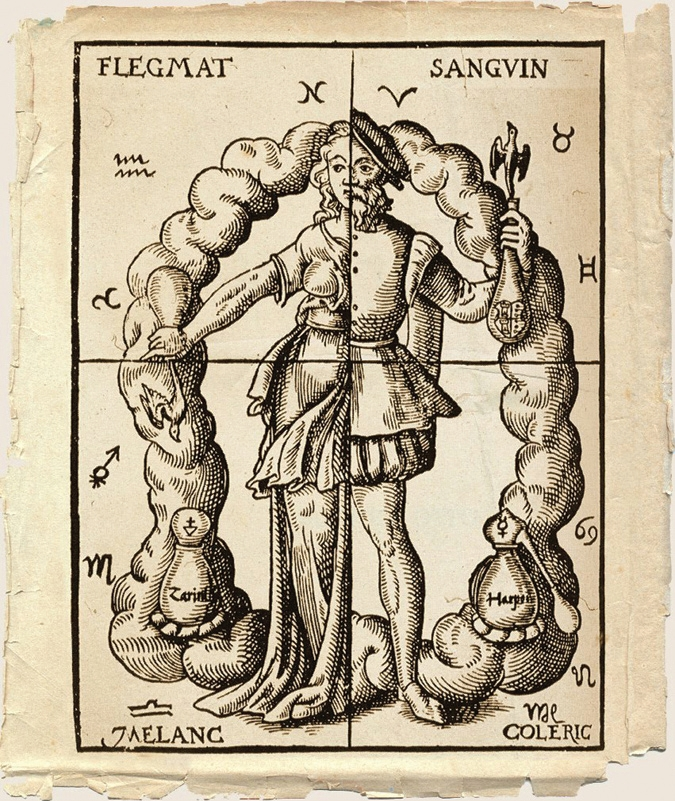
    Beyond naming the shapes of tumors, Hippocrates and Galen also formulated an influential theory to explain what caused the crab to grow inside the body. Operating under the ancient humoral theory, which held that human health depended on the balance of four bodily fluids—blood, phlegm, yellow bile, and black bile—Galen argued that cancer was caused by a dangerous localized trapping and thickening of atra bilis, or black bile. Because black bile was considered dark, cold, and acidic, Galen taught that when the spleen failed to clear excess black bile from the bloodstream, the dark fluid settled in tissues like the breast or stomach and solidified into a crab like tumor, a medical belief that dominated European universities for over fourteen hundred years until the rise of cellular pathology in the nineteenth century.

  • History of Cancer

    History of Cancer

    Before the Common Era

    Traces of Cancer in the Remains of Ancient People

    In 1990, during excavations of the Chiribaya tribe cemetery in Peru on the northern border of the Atacama Desert, American professor Arthur Aufderheide discovered the mummy of a young woman with osteosarcoma — a malignant bone tumor. These remains were well-preserved thanks to the local climate: clay extracted all the liquid from the body, and the desert wind dried the tissues. However, scientists do not always find remains of tumor tissues. Sometimes, they come across traces of the disease left in the body of an ancient person, such as small holes in the skull and shoulder bones — results of metastases from skin melanoma or breast cancer.

    Incurable “Lumps in the Breast” from the Edwin Smith Papyrus

    One of the earliest written records of the disease is the Edwin Smith Papyrus, named after the American archaeologist who bought the artifact in an Egyptian market in 1862. This ancient Egyptian manuscript, dated to around 1600 BCE, is likely an incomplete copy of an earlier medical treatise created in the 27th century BCE. Its authorship is attributed to the famous Imhotep — an architect and healer of the Old Kingdom period. The surviving document describes 48 cases of various injuries, with the forty-fifth section unexpectedly devoted to the consequences of a severe specific disease — presumably breast cancer:

    When you examine the swelling lumps in the breast and find that they have spread throughout the breast, and if you place your hands on the breast, you find no heat and the tissues feel cool, with no graininess, internal fluid, or liquid discharge, but they protrude when touched, then you can say of the patient: ‘Now I treat the growth of tissues… the enlarged lumps of the breast mean the presence of swellings in the breast, large, spreading, and firm, and touching them is like feeling a ball of bandages, or they may be compared to an unripe fruit, hard and cool to the touch…’.

    For each injury described in the papyrus, the author suggests different treatments: for example, poultices for wounds and balms for burns. However, the mysterious “swelling lumps in the breast” baffled the ancient physician — unable to find a cure for this ailment, the “Treatment” section dryly states: “None exists.”

    The Persian Queen Atossa and the Successful Surgical Treatment of Breast Cancer from Herodotus’ “Histories”

    Two millennia after the Imhotep Papyrus, a similar disease reappears in written sources. Herodotus, in his “Histories,” tells of the Persian queen Atossa, who suffered from a bleeding breast tumor. Some researchers believe she had inflammatory breast cancer. None of the remedies helped Atossa, so the Greek physician Democedes proposed surgically removing the malignant tumor. The queen agreed, and the operation ultimately saved her life. Thus, Atossa’s story can be considered the first recorded example of a successful mastectomy — a surgical procedure to remove the breast.

    Ancient Rome

    Black Bile

    The four humors. Illustration from Leonard Tourneisser's book Quintessence. 1570
    The four humors. Illustration from Leonard Tourneisser’s book Quintessence. 1570

    Roman physician Galen, who lived from 129 to 216 CE, following Hippocrates, believed that a healthy person’s body was in balance with four humors (fluids): blood, phlegm, yellow bile, and black bile. An excess of any humor inevitably led to illness, and treatment involved removing excess fluid from the body, for example, through bloodletting, emetics, and laxatives.

    Galen believed that malignant tumors were caused by an excess of black bile, which became a dense mass in the body. Interestingly, medieval physicians believed that an excess of black bile also caused melancholy: μελαγχολία in ancient Greek means “black bile.” Galen, whose authority remained unchallenged for over a thousand years, argued that the disease could not be defeated by surgery: the black bile would remain and pose a threat of new tumors. According to him, a patient could only be supported with general therapeutic measures.

    Middle Ages

    Boar Tusk, Fox Lungs, and Ground Elephant Bone

    Galen’s views predetermined the medieval medical approach to tumors. Surgery was generally considered more harmful than beneficial, and the absence of radical treatment was seen as the best possible remedy. An alternative to the surgeon’s knife was a wide range of rather exotic remedies: arsenic tincture, lead tincture, boar tusk, fox lungs, ground elephant bone, crushed white coral, castor bean seeds, senna plant, and more. Alcohol and opium tincture were used to relieve unbearable pain.

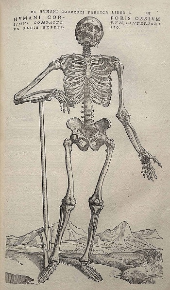
    Image from Andreas Vesalius' De humani corporis fabrica (1543), page 163
    Image from Andreas Vesalius’ De humani corporis fabrica (1543), page 163

    In the Renaissance, medicine revisited Galen’s idea that an excess of black bile in the body caused malignant tumors. Andreas Vesalius, the founder of scientific anatomy and author of “On the Fabric of the Human Body in Seven Books” (1543), who personally dissected corpses, could not find even the slightest trace of the infamous substance supposedly responsible for the development of tumors.

    Enlightenment: Cancer from an Excess of Lymph

    Another suspected cause of malignant tumors was lymph (also known as phlegm in humoral theory). According to this new understanding, tumors were no longer attributed to mysterious black bile but to a clear fluid that permeates the human body and is responsible for a calm temperament. German physicians of the 17th–18th centuries, Georg Ernst Stahl and Friedrich Hoffmann, suggested that tumors consisted of fermented lymph, which could have different densities, acidities, and alkalinities in each specific case.

    Cancer as Contagion

    While Hippocrates, Galen, Stahl, and Hoffmann sought the cause of the disease within the body, two 17th-century Dutch physicians, Zacutus Lusitanus and Nicolaas Tulp, found it outside. Independently, they reached the same conclusion: cancer is contagious. The doctors proposed moving all patients outside major cities to isolate them and thus prevent the spread of the dangerous disease. The idea of the contagiousness of cancer, prevalent in Europe during the 17th and 18th centuries, is now considered mistaken. However, it is known that certain viruses, bacteria, and parasites can indeed increase the risk of developing tumors, such as the human papillomavirus or the bacterium Helicobacter pylori.

    Cancer from Lifestyle

    In the 18th century, scientists made three important discoveries that laid the foundation for the epidemiology of cancer. In 1713, Italian physician Bernardino Ramazzini noted that cervical cancer was almost nonexistent among nuns, while the incidence of breast cancer was relatively high. Ramazzini concluded that lifestyle (in the case of nuns — lack of sexual relations) could directly influence the development of this disease in women.

    In 1775, English surgeon Percivall Pott discovered that scrotal cancer, often found in chimney sweeps, had an occupational nature: tumors arose due to the accumulation of soot in the folds of the scrotum. Pott’s discovery later led to the study of carcinogens associated with specific professions and the gradual adoption of occupational health and safety measures.

    By the 20th century, it was established that carcinogens could include benzene, used in the production of medicines, plastics, and rubber, as well as the fine-fiber mineral asbestos, used in construction, among others.

    It also became clear in the 18th century that bad habits could cause this disease. In 1761, English scientist John Hill, in his book “Cautions against the Immoderate Use of Snuff…,” first directly linked tobacco use and the development of tumors. However, lung cancer caused by smoking only became a subject of intensive research in the 20th century.

    Surgery as the Only Way to Defeat Cancer

    The medicine of the Enlightenment era drew a final line under Galen’s teachings. While Vesalius created a scheme of the “healthy” human body, the English anatomist Matthew Baillie managed to describe the “pathological” body in minute detail. In his work “The Morbid Anatomy of Some of the Most Important Parts of the Human Body” (1793), Baillie thoroughly examined various malignant formations, but in none of them could he find any hint of black bile.

    Having “buried” black bile as a scientific error, medicine paved the way for surgery as possibly the only effective method of fighting malignant tumors. However, as before, patients could only rely on the skill of doctors. Indeed, in the absence of means to alleviate their condition during operations, it was difficult to count on a favorable outcome: people who agreed to go under the surgeon’s knife typically died from pain shock, heavy blood loss, and various infections.

    19th Century

    Ether and Carbolic Acid

    The situation changed dramatically in the middle to late 19th century with the invention of anesthesia and antiseptics. American dentist William Morton began using the organic substance ether as a general anesthetic, while English surgeon Joseph Lister started using carbolic acid to disinfect wounds. After these important discoveries, doctors were able to resort to radical surgery when it was necessary to remove a tumor along with lymph nodes.

    Surgeons such as Theodor Billroth and William Stewart Halsted became famous in this field. Billroth performed the first esophagectomy (removal of part of the esophagus), laryngectomy (removal of the larynx), and gastrectomy (removal of the stomach) in history, while Halsted performed radical mastectomy, which is the complete removal of breast tissue.

    Cell Theory

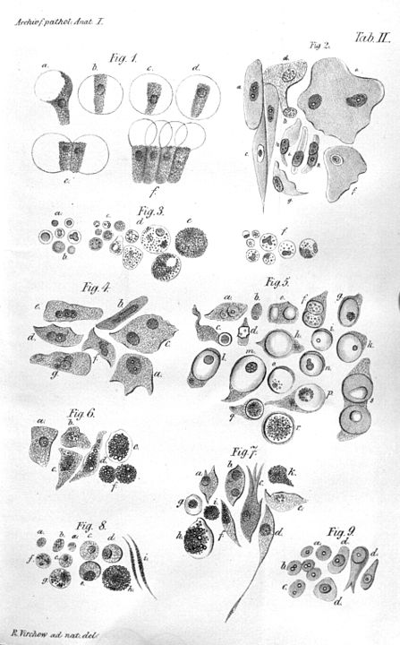
    Illustration of Virchow's cell theory
    Illustration of Virchow’s cell theory

    In the 19th century, as microscope design was improved, scientists began to gradually approach an understanding of the true nature of cancer. In 1838, German biologist Johannes Peter Müller, in his work “On the Finer Structure and Form of Morbid Tumors,” proved that malignant formations consist not of lymph but of cells. However, he still believed that tumor cells were formed not from normal cells but from blastema between them. But his student Rudolf Virchow proved that all cells, including tumor cells, are formed from other similar cells: omnis cellula e cellula (“all cells come from cells”).

    However, even Virchow was mistaken: he was sure that cancer spread through the body like a fluid. In the 1860s, German surgeon Karl Thiersch disproved Virchow, proving that tumors consist of epithelial tissue, not connective tissue. He showed that metastases appear as a result of the spread of malignant cells, not through some unknown fluid.

    20th Century

    Hormone Therapy

    In the late 1870s, English physician George Thomas Beatson discovered a direct connection between the ovaries and milk production in the breast: after removing this organ in rabbits, he noticed that they stopped producing milk. This discovery led Beatson to wonder: could oophorectomy, or removal of the ovaries, have a positive effect on advanced breast cancer? Indeed, his experiments showed that such an operation often helped improve the condition of women with this type of cancer.

    The scientist also suggested that the ovaries themselves might be the main cause of breast cancer development. Even before estrogen was discovered, Beatson determined that the female hormone from the ovaries had a stimulating effect on breast cancer. Beatson’s discoveries laid the foundation for modern hormone therapy, where substances like tamoxifen, which suppresses the effects of estrogen, and aromatase inhibitors, which inhibit enzymes that convert male hormones (androgens) into female hormones (estrogens), are used to treat and prevent breast cancer.

    In the 20th century, a hormonal method was also found to treat a male disease—prostate cancer. In the 1940s, American scientist Charles Huggins discovered that after castration, patients experienced a sharp regression of metastatic prostate cancer. In addition, it was found that simultaneously decreasing testosterone levels and increasing estrogen levels helps in treating “male” cancer.

    Radiation Therapy

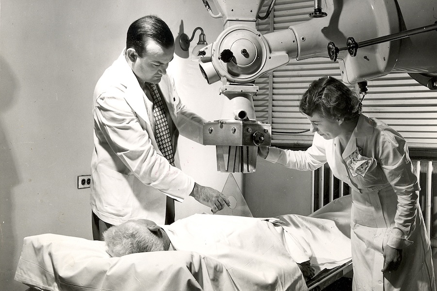
    Radiation therapy at MSK circa 1949
    Radiation therapy at MSK circa 1949. Image: Memorial Sloan Kettering Cancer Center

    At the end of the 19th century, Wilhelm Röntgen discovered X-rays, and Marie Curie and Pierre Curie discovered radium. Along with this came a new direction in tumor treatment: radiation therapy. Scientists found that radium could damage tumor cells in the body to the point of their complete destruction. However, in the early stages of radiation therapy, many doctors did not fully realize that radiation attacks healthy cells just as successfully as diseased ones. If the necessary dose is miscalculated, the rays can be deadly.

    It took almost a century to fully control unmanageable radiation. At the end of the 20th century, conformal radiation therapy was invented, in which the beam is precisely directed at tumor tissues thanks to detailed three-dimensional models created using computed tomography.

    Chemotherapy

    The militarism of World War II killed millions of human lives but unexpectedly helped find a new means in the fight against malignant tumors. In the 1940s, as part of developing more effective weapons, American scientists Louis Goodman and Alfred Gilman, commissioned by the US Department of Defense, studied chemical substances related to mustard gas, a poisonous chemical compound. During these studies, they accidentally found that nitrogen mustard helps in treating cancer of the lymph nodes (lymphoma). This was one of the first steps towards introducing chemotherapy into medical practice.

    Sidney Farber. 1960
    Sidney Farber, 1960. Image: Public Domain

    The real era of chemotherapy began with Sidney Farber, an American oncologist who, in the late 1940s, proved that a substance called aminopterin could cause remission in children suffering from acute leukemia: it blocks the division of leukocytes. Subsequently, adjuvant therapy became widely used in medical practice, which is a special chemotherapy aimed at destroying all tumor cells remaining in the body after surgery. Such therapy was first tested in the treatment of breast cancer, then in the treatment of colon cancer, testicular cancer, and other diseases. Another important innovation was combination chemotherapy, in which several different drugs are used simultaneously for more effective treatment.

    Immunotherapy and Targeted Drugs

    One of the most effective modern means in the fight against malignant tumors is immunotherapy. It has been actively used since the 1970s. Immunotherapy involves introducing special biological agents into the patient’s body that can both mimic the natural immune response and help the body’s own immune cells fight the tumor. The first targeted drugs—rituximab and trastuzumab—were created in the late 1990s; since then, they have been used to treat lymphoma and breast cancer, respectively.

    Lymph node with mantle cell lymphoma
    Lymph node with mantle cell lymphoma. Image: Gabriel Caponetti, CC BY-SA 3.0

    A promising direction in modern immunotherapy is the development of special cancer vaccines. For example, in 2010, the first vaccine was approved in the USA for a form of prostate cancer that no longer responds to hormone treatment. This drug, unfortunately, cannot kill the disease, but it helps the immune system fight tumor cells, prolonging the life of patients who seem to have a fatal diagnosis.

    Another important direction in modern immuno-oncology is the development of checkpoint inhibitors that regulate mechanisms that block the body’s immune response. The first of these drugs, ipilimumab, was approved in the USA in 2011. Now patients with diseases that were previously considered incurable have a chance for effective therapy. For example, former US President Jimmy Carter was able to get rid of metastatic melanoma when he was already over 90 years old.

    Also, quite recently, cell therapy using lymphocytes taken from the patient, which are then genetically modified, began to spread. It was first and successfully used against acute lymphoblastic leukemia in a five-year-old American girl, Emily Whitehead. In addition, research in the field of gene therapy continues. In general, modern medicine strives for ever-greater personalization of treatment, which makes it possible to completely cure the disease.

  • Why Penile Cancer Cases Are Increasing?

    Why Penile Cancer Cases Are Increasing?

    Although penile cancer remains a relatively rare malignancy compared to prostate, lung, or colorectal cancers, recent global epidemiological studies have revealed a concerning upward trajectory in incidence rates across both developing nations and high income European countries. Data compiled by international cancer registries indicate that global diagnoses are projected to climb by more than seventy percent by the year 2050. Understanding why this once obscure urological condition is rising requires examining the intersection of shifting demographic pyramids, persistent viral infections, changing lifestyle habits, and historic disparities in preventive hygiene and vaccination.

    The Role of the Aging Global Population

    The single largest statistical driver behind the projected surge in penile cancer cases is the rapid aging of the global male population. Penile squamous cell carcinoma, which accounts for roughly ninety five percent of all malignancies in this anatomical region, is overwhelmingly a disease of older adulthood, with the vast majority of diagnoses occurring in men over the age of sixty. Because cellular DNA repair mechanisms naturally lose efficiency over decades of life, chronic tissue inflammation and latent viral infections accumulated in youth often take thirty to四十 years to trigger malignant cellular transformation.

    As life expectancy increases across South America, Asia, and Africa, and as the large post war baby boomer generation moves into its seventies and eighties across Europe and North America, the sheer number of men living within the peak age bracket for urological malignancies has expanded dramatically. Even in regions where age standardized risk per individual has crept upward only slightly, the demographic shift toward an older citizenry guarantees a steep rise in absolute hospital caseloads over the coming decades.

    Human Papillomavirus and Changing Sexual Patterns

    Beyond aging demographics, the primary biological engine driving new cases in younger and middle aged men is infection with high risk strains of the Human Papillomavirus, particularly HPV 16 and HPV 18. Pathological analyses demonstrate that approximately half of all penile cancer tumors worldwide contain integrated HPV viral DNA. When the virus infects the mucosal epithelium of the glans or foreskin, it produces oncoproteins known as E6 and E7, which bind to and deactivate the human body’s natural tumor suppressor proteins, p53 and retinoblastoma, allowing damaged cells to divide uncontrollably.

    Epidemiologists tracking rising cancer rates in countries such as the United Kingdom, Germany, and Scandinavia point to shifts in sexual behavior that began in the late twentieth century. Higher numbers of lifetime sexual partners, earlier ages of first intercourse, and inconsistent barrier protection have increased the circulation of high risk HPV strains across the general population. While female cervical cancer rates have begun to plummet in wealthy nations thanks to decades of routine Pap smear screening and early targeted HPV vaccination campaigns for girls, boys were initially excluded from many national vaccination rollouts, leaving a substantial cohort of adult men unprotected against oncogenic viral strains.

    Phimosis, Chronic Inflammation, and Declining Circumcision Rates

    The second major pathway to penile malignancy is entirely independent of viral infection and instead stems from chronic, long term mechanical and chemical irritation of the skin. Men with phimosis, a condition in which a tight foreskin cannot be fully retracted over the glans, face a markedly elevated risk of developing cancer—sometimes up to ten times higher than men without the condition. When the foreskin cannot be retracted cleanly, cellular debris, natural oils, and moisture accumulate underneath to form smegma, creating an environment prone to bacterial colonization and repetitive inflammatory micro trauma.

    Related dermatological conditions, such as lichen sclerosus (balanitis xerotica obliterans), cause chronic scarring and white patchy atrophy of the genital tissue that frequently progresses into precancerous lesions if left untreated with topical therapies. Historically, neonatal circumcision nearly eliminated the risk of phimosis related penile cancer by removing the tissue most susceptible to chronic subpreputial inflammation. In several Western nations where routine infant circumcision rates have declined over the past forty years without corresponding public health education on foreskin hygiene and early medical treatment for phimosis, urologists have observed a parallel rise in inflammatory squamous cell lesions.

    Smoking, Socioeconomic Barriers, and the Path to Prevention

    Lifestyle cofactors further accelerate the progression from mild tissue dysplasia to invasive carcinoma. Tobacco use—both cigarette smoking and smokeless chewing tobacco—exposes the entire urinary and reproductive tract to carcinogenic nitrosamines and polycyclic aromatic hydrocarbons excreted through urine and sweat, while simultaneously suppressing the local Langerhans immune cells responsible for clearing HPV infections from the skin. Studies consistently show that heavy smokers face more than double the risk of developing invasive penile cancer compared to non smokers.

    Compounding the biological risk is a powerful psychological barrier: embarrassment and social stigma. Across every continent, men frequently ignore early warning signs—such as painless persistent red rashes, small crusty ulcers, warty growths, or changes in skin color beneath the foreskin—for more than a year before consulting a physician, often mistaking early tumors for harmless chafing or sexually transmitted infections. Reversing the global rise in penile cancer depends on expanding gender neutral HPV vaccination to boys worldwide, normalizing routine urological self examinations, treating phimosis early in life, and dismantling the silence that prevents men from seeking timely medical care.

  • Viruses and cancer: Discovery of an “impossible” relationship

    Cancer is a terrifying pandemic that hangs over our heads like the sword of Damocles. Innumerable individuals are afflicted by this multifaceted illness, and tens of thousands more must live with the diagnosis every day. Even with the best diagnostic tools and treatment options available, many people still die from it. Doctors are working tirelessly to determine what has triggered this cancerous multiplication of cells.

    Some of the possible causes of cancer have been identified as exposure to toxins or radiation or a family history of the illness, although unlike with many infectious diseases, a direct correlation between cause and effect is usually not sufficient for cancer. The catastrophic transition from a healthy body cell to a tumorous cancer cell is often set in motion by the confluence of a number of elements. However, scientists have barely scratched the surface of this interaction’s intricate complexity.

    Tumor viruses are one aspect that has gained the attention of scientists. The earliest signs that viruses may cause cancer were identified by scientists around the turn of the past century, but this information was largely disregarded for decades. In the past, there was no link between infectious agents and tumor diseases.

    Despite the fact that the link between cancer and viruses was finally proven, scientists in this area faced an unprecedented wall of opposition. Ignorance, but also tenacity, has a long and illustrious history that continues up to the present day. As such, it served as a near-perfect illustration of the typical sequence of scientific progress described by Austrian engineer and author Robert Musil: “Every few years that something till then held to be in error suddenly revolutionizes the field, or that some dim and disdained idea becomes the ruler of a new realm of thought.”

    More and more viruses are being added to the list of “suspects” by cancer researchers, and roughly a dozen have been recognized beyond question, demonstrating the “new realm’s” visible growth. The human papillomavirus and various herpes viruses are two examples of these ubiquitous yet purportedly innocuous infections. Viruses’ causal pathways in cancer are very vaguely understood, but researchers are working to fill up the gaps.

    The discovery of the cancer virus

    Chickens to revolutionize science

    In the year 1909, a farmer pays a visit to a young physician and biologist Peyton Rous at his new job at New York’s Rockefeller Institute for Medical Research. He is concealing a jar with a cancerous chicken under his arm. A sarcoma, a huge cancerous tumor of connective tissue, had developed on the right side of the breast of this striped Plymouth Rock chicken.

    At the time, research on cancer was still in its infancy, but Rous, who worked on the subject, jumped at the chance to do a battery of experiments using the chick and its tumor. So, he decided to produce an extract from the diseased tissue, filter it to remove any remaining cells, and then inject it into young chickens of the same breed. Unusually for the period, the healthy animals also got cancerous tumors of the same sort as a consequence of this experiment.

    With this in mind, Rous deduced that the tumor was caused by a microscopic pathogen present in the extract. He suspected a parasitic creature, probably one of the recently identified viruses. Rous attempted to prove his hypothesis by successfully transmitting additional chicken cancers via this route. His findings and theorization of a cancer-causing virus were published in 1910, setting off a real “tumor wave.”

    Standing on the sidelines

    However, no success was found in using this method to induce growth in any other animal species. Soon after, Rous was marginalized in the scientific community, and his viral tumors were deemed to be a chicken-specific anomaly. In his acceptance address for the Nobel Prize in 1966, Rous recalled, “The results of the sarcoma virus were met with absolute disbelief.” However, “spontaneous” chicken tumors with different morphologies were successfully transplanted and a virus was found in each of them not long after.

    In the 1930s, Richard Shope, a colleague of Rous’s, obtained comparable results using an extract of a benign skin tumor, a papilloma, from the skin of a wild rabbit species. However, these findings were disregarded since they were not thought to be relevant to cancer.

    “Milk factor” instead of viruses

    The standard medical view, that cancer was at most related to a person’s genes or their environment and could not be caused by viruses or other pathogens, appeared to be impervious to new evidence. Under the banner of “What must not be, cannot be,” any and all theories involving viruses that cause cancer were dismissed as ridiculous. In the 1930s, the United States’ highest health authority convened a council of specialists who issued a formal guiding principle that viruses and other organisms could be ruled out as causes of cancer.

    Due to the atmosphere, few scientists were willing to publicly provide proof of the presence of cancer viruses. Breast cancer-causing viruses were first identified in mice by John Bittner, a researcher at the leading cancer research facility in the United States, in the late 1930s. Instead of calling the cancerous agent a virus in his paper, he called it a “milk factor,” neutrally avoiding the risk of losing his job and future research funding.

    The breakthrough

    The significant change, and the subsequent rise of cancer virus researchers, did not come until the late 1950s. There were two primary causes that resulted in this change: Scientists found that some viruses could introduce their genetic material into the host cell’s DNA. This modified the cells irreversibly without killing them. However, numerous scientists started to find tumor viruses in their lab tests, making it impossible to deny the undesirable reality.

    According to a long-held theory, cancer and infections produced by viruses or other organisms could not be linked to one another. But a decade later, in 1966, Peyton Rous was honored for his foresight and tenacity with the Nobel Prize in Medicine, and the virus he discovered was given the honorific name “Rous sarcoma virus.“

    Looking for the virus-cancer mechanism

    Genome thieves

    Cancer cells are the progenitors of more cancer cells and pass on their destructive capacity to their children, making them the ultimate misfits. Yet they originate in healthy tissue. But, how? What part do viruses and other pathogens play in this process? Caltech cancer researcher Renato Dulbecco was pondering this very subject. As a result of Peyton Rous’s rehabilitation, study into tumor viruses is no longer taboo, although still very little is known about how they work.

    In 1960, he and his colleague Marguerite Vogt undertook laboratory experiments in which they infected hamster cells with a polyoma virus, a DNA virus with little complexity. The cells started to transform as expected, and tumors started to form. However, they immediately noticed something peculiar: the viral reproduction ceased along with the transition to cancer cells. Their numbers did not increase or decrease as they would during an actual infection since they were not able to exhaust the cell’s resources on viral production. Sure, but why?

    The virus always leaves behind traces

    Dulbecco could only think of two possibilities: either the virus got killed out in the process of the cell’s metamorphosis, or it survived the transformation but was unable to multiply. After much effort and testing, the scientist finally got an answer. He discovered the virus’s “footprints” in the changed cells, disproving the original hypothesis and confirming the infection was still there.

    Using molecular biology techniques, he was also able to show that the virus was inactive since a key piece of its DNA had been incorporated into the host cell’s genome, rendering it inaccessible for viral replication. Only around seven proteins were encoded by the viral DNA that was added, but that seemed to be enough for cell processes to go haywire.

    Contrary to dogma

    A young scientist at the University of Wisconsin named Howard Temin had less luck than Dulbecco did when he published his findings and received both praise and criticism for them. Unlike Dulbecco, Temin studied an RNA virus (the Rous sarcoma virus) in his tests; yet, he reached the same conclusion as Dulbecco in a 1964 paper: the virus must add something to the cell’s genome that initiates the cell alterations. According to him, before inserting its genetic material into cellular DNA, the virus was “translating” its RNA genome into DNA, called the “provirus.”

    However, he made a huge “error” by proposing this theory, since it challenged the prevailing paradigm in molecular biology at the time. This dogma states that information in live cells always flows from DNA to RNA and then to protein, but never the other way around. For this reason, most of Temin’s scientific peers would have considered his concept of a provirus that employed this “wrong” path to be heretical. The response was just as furious and condescending.

    Again, it took almost a decade and the “administrative aid” of a coworker before the previously disgraced scientist was vindicated. Temin and the biologist David Baltimore delivered the killing blow to the doctrine at the decade’s close in the 1970s. Both separately found an enzyme in tumor virus-infected cells that can convert RNA to DNA. There was experimental proof that this “provirus DNA” was integrated into the cellular genome.

    The seminal work on the biology of tumor viruses by Dulbecco, Temin, and Baltimore was recognized with the Nobel Prize in 1975. However, the three of them were unable to resolve the lingering mysteries of how viruses cause cancer and whether or not they also cause cancer in humans. “I do not believe that infectious viruses are the causes of most human tumors, but I think that viruses can provide us with models for the development of cancer,” Temin said cautiously in his Nobel Prize acceptance address.

    Looking for a cancer gene

    Can cancer viruses replicate without causing disease?

    Two researchers at the U.S. National Cancer Institute, Robert Huebner and George Todaro, experimented with the Rous virus on various mammals in the 1970s, shocking the scientific community just as they were coming to terms with the discovery that cancer viruses, including and especially RNA viruses, incorporated part of their genetic information into the DNA of the cell. They found, to their shock, that viral DNA could be detected in tumor cells even when the tumors had not been caused by the virus but rather by physical or chemical effects. How could this be explained?

    The “virogen-oncogene hypothesis”

    Huebner and Todaro proposed that viral DNA doesn’t come “out of nowhere” since cells must already have viral genes embedded in their genetic material, which were incorporated into the genome at some point during evolution. These conserved DNA regions are inactive under normal conditions but become functional when exposed to certain stimuli, allowing them to produce viral proteins and, in turn, new viruses. However, they postulated that some of these “virogens” also operated as “oncogenes,” causing the development of tumors as well.

    Hunting down the tumor gene

    If this hypothesis were to hold, then the Rous oncogene must also be present in the cellular DNA of normally developing animals. Michael Bishop and Harold Varmus of the University of California, San Francisco, began a mission to match the genome of the Rous sarcoma virus with chicken DNA for this very aim. But this would be very difficult to do without high-tech equipment like PCR or sequencing devices.

    Fortunately, in 1971, a serendipitous discovery by Peter Vogt of the University of Southern California revealed a viral mutant that was capable of replication but seemed to lack the potential to produce malignant tumors. The virus’s genome was 15 percent shorter than average, suggesting that this missing part could include the critical tumor gene(s).

    This was the key piece of information that helped Bishop and Varmus solve the case. This allowed them to determine whether or not the “src” gene fragment was present in the cellular DNA of their guinea pigs and, if so, whether or not it functioned as a possible cancer gene. However, scientists were surprised to discover that the virus was present not just in chickens, the primary hosts of the Rous virus, but in birds of every genus and subgenus.

    But that wasn’t the end of it; other researchers soon identified src variations in guinea pigs, rabbits, rats, snakes, and even humans. The oncogene postulated by Huebner and Todaro seemed to be found. Bishop and Varmus, though, were not confident in the claims. They were wary about its extraordinarily widespread distribution. The origin of the gene still remained unknown; did this gene really come from the Rous virus?

    Oncogenes: a biological enigma

    The enemy within

    An essential step forward accomplished with the discovery of a cellular proto-oncogene. Not much time had passed, yet scientists had already uncovered over 40 distinct retroviral oncogenes. They speculated that the majority of cells had gene sequences that were homologous to a viral gene known to cause tumors. Tumor viruses and other external factors could contribute to tumor development, but there was also an “internal adversary” that might be just as crucial.

    However, more questions were created by the new data than they answered. Among the most crucial, in Varmus’s words, “Did the evolutionary conservation of the src gene indicate its cellular origin? Or could it still be a virus-derived gene that was just better conserved than comparable viral elements?”

    Own cell instead of being smuggled in

    Rapid responses to this question emerged from many different fields. Sequence analysis, however, provided the clinching evidence. It demonstrated that the src gene was broken up into many segments by introns (“junk DNA”), which were typical of biological DNA but were rarely seen in viral genes.

    Harold Varmus reflected on this, saying, “All our arguments and experiments led to only one conclusion: the precursor to src was a conserved – and therefore vital – cellular gene that found its way into the Rous sarcoma virus by recombination.” So, it seemed that the proposed evolutionary route of the “virogen oncogene” concept must have followed an exactly opposite direction.

    Since the oncogene’s precursors have been conserved throughout evolution, this explained why they are found in so many different types of cells. “Far from being a harmful element just waiting to be activated by a carcinogenic signal, the precursors of viral oncogenes seem to have an impact on the organism to have an important function, otherwise they would not have been preserved in the course of evolution,” said Varmus. These genes are, in fact, involved in essential cellular processes, including multiplication and differentiation.

    Hostile takeover

    How, therefore, could this normally helpful gene cause an infected cell to go berserk? Bishop and Varmus hypothesized that the retrovirus identifies the benign proto-oncogene during infection and alters it into the malignant oncogene. During this process, known as transduction, the virus inhibits the gene’s regular function and removes it from the organism’s regulatory system. Therefore, the misplaced and overactive genes have undesirable effects, such as excessive cell multiplication.

    The two scientists speculated that Huebner and Todaro’s peculiar results might be explained if the transition was triggered by chemical or physical forces. In 1983, researchers found a group of cellular genes that were structurally similar to viral cancer genes and oncogenes from non-viral cancers, lending credence to this theory.

    Viruses as a possible cause of human cancer

    Initial evidence for human T-virus type 1

    By pinpointing the “inner enemy” and showing that viruses might play a role as triggers in carcinogenesis, Bishop and Varmus have thrown open the floodgates for contemporary cancer research. They shared the Nobel Prize in Medicine in 1989.

    However, they encountered resistance and even contempt for their ideas. Their hypothesis, that a comparable process could possibly contribute to human tumor formation, was at odds with the current scientific consensus. But this perspective held that although viruses were identified as a cause of tumor disorders in animals, there was less evidence to support a similar link in humans. But they weren’t completely wrong since hereditary or environmental factors are still considered to be the main causes of cancer in humans.

    The first human retrovirus

    However, this started to change in the early 1980s. In 1980, a patient with cutaneous T-cell lymphoma, a benign skin ulcer, was investigated by virus researcher Robert Gallo and his colleagues at the National Cancer Institute in the United States. He analyzed the tissue samples and found a retrovirus, a kind of virus that had never been seen in humans. While these RNA viruses, to which the Rous sarcoma virus also belongs, had been studied for quite some time in animals, this was the first time that they had been found in people.

    Gallo and his Japanese colleagues continued studying its biology and ecology in the years that followed. Scientists found an unexpected link between the virus and adult T-cell leukemia lymphoma; whereas most infected people had relatively mild skin changes over time, a small percentage had acquired this aggressive type of leukemia.

    This kind of cancer often does not manifest until the age of 60, but once it does, it generally results in a swift death. It is estimated that just 1–4% of infected people actually acquire leukemia, but all patients carry the virus anyway.

    And the Leukemia

    So, was the virus the cause of cancer? There had to be more evidence than just finding a virus in a cancer cell. But in 1982, Gallo and his group found the viral “culprit” in the virus’s DNA. Since there was no homolog of the viral gene “tax” in the human cell genome, “tax” could not be considered an oncogene in the traditional sense. But when it was inserted into the cell’s DNA, it activated particular T cell growth factors, leading to the unchecked multiplication characteristic of leukemia. The specifics of this mechanism were, however, still little known.

    The pathogen, now known as HTLV-1, or human T-cell leukemia virus, made medical history as the first virus known to cause cancer in humans. This discovery sparked what has become a decades-long effort to identify and characterize additional viruses that might cause human tumors. Even while the hepatitis C pathogen was now thought to have a role in certain kinds of liver cancer and maybe even in some forms of non-lymphoma, Hodgkin’s other RNA viruses had thus far been on the suspect list at best, with nothing yet confirmed.

    Human papillomavirus (HPV) and cervical cancer

    Forming a model

    Human papillomavirus (HPV) is a tiny, contagious virus that may manifest in anyone’s cells. The current global prevalence of this DNA viral infection is estimated to be above 25 million. The sexually transmitted disease affects almost half of the male and female population.

    Infected people may not even realize they have it. The virus is asymptomatic and may live in the skin and mucous membranes for a long time; in rare instances, benign warts may appear. In addition to its pervasiveness, HPV is also remarkably diverse, with over 130 distinct strains having been found to date. And 18 of them are particularly worrisome since they belong to high-risk categories and may cause cancer. Discovered in 1983, this link between a virus and cancer is now one of the best studied and proven anywhere. Some virologists think that HPV is responsible for at least 600,000 instances of cancer globally; other estimates place the number at between 12 and 15 percent of all tumor cases.

    Two proteins that sneak into cells

    The human papillomavirus types 16, and 18, are mostly to blame. More than 90% of head and neck cancers and 99% of cervical cancers have these abnormalities. In most cases, the human immune system can clear up an HPV infection in around ten months. Changes occur, however, when risk factors are introduced, such as smoking, an impaired immune system, or a particularly aggressive viral strain:

    Two viral proteins, E6 and E7, are produced by the infected cells. When bound to a particular cell protein, E6 triggers that protein to label the tumor suppressor gene P53 for destruction. Consequently, the cell is deficient in a key component for preventing mutations and cancerous transformations. Another viral protein, E7, is responsible for cell death by “hijacking” a cellular protein engaged in crucial regulatory mechanisms. This causes the release of transcription factors that actively promote cell multiplication. Two proteins assist the virus in disrupting the cell’s basic metabolic and developmental processes, leaving the cell vulnerable to mutations.

    However, this is by no means the sole mechanism by which HPV causes cervical cancer. In fact, new mechanisms are disclosed virtually every week. These two, though, are the sharpest and most consequential.

    The dirty dozen

    Researchers have had a hard time finding new tumor-inducing viruses due to the intricacy and variety of the processes and regulatory systems already engaged. This is one of the reasons why just a handful of anti-cancer drugs that target viruses have been shown to be effective in people.

    Epstein-Barr virus (EBV) for nasopharyngeal cancer and Hodgkin’s lymphoma; hepatitis viruses B and C for liver cancer; human T-cell lymphotropic virus type 1 (HTLV-1) for cervical cancer; and human herpesvirus 8 (HHV-8) as a trigger for Kaposi’s sarcoma are all well-established viruses that cause cancer in humans. Polyomavirus and hepatitis C for non-Hodgkin’s lymphoma, and an unidentified agent for a kind of pediatric leukemia, remain on the list of possible tumor-causing viruses. The tally of cancer viruses in humans that have been “condemned” continues to increase today.

    Helicobacter pylori and stomach cancer

    Several kinds of cancer in humans now have a known link to infections with certain viruses, bacteria, and parasites. 15–20% of all malignancies are believed to have an infectious cause. But we weren’t always aware of the cancer-causing bacteria. In fact, this medical dogma was toppled anew by a dogged scientist, a little bit of self-experimentation, and a giant leap forward. The Australian scientist Barry Marshall, his colleague Robin Warren, and the rod-shaped bacteria Helicobacter pylori are the story’s primary characters.

    In 1982, the medical community accepted the dictum that “Bacteria are everywhere—but the human stomach is bacteria-free.” Because of the very acidic conditions present in the stomach, it was often believed that no bacteria could thrive there. And that was not all. The majority of medical professionals and scientists in the field of pharmacology, as well as patients, attributed the pain of gastric mucosal inflammation and subsequent gastric ulcers to the presence of acid in the stomach. The pharmaceutical industry promoted acid blockers of different sorts with the slogan “No acid, no ulcer,” and sales were booming as a result.

    Its home is the stomach

    Pathologist Robin Warren found something that shouldn’t be there when he examined tissue samples from stomach ulcers and irritated stomach mucosa under a microscope: bacteria that are full of mucus. And that was the case in over half of the samples he tested. The number of these cells seemed to be correlated with the degree of inflammation. Warren consulted with doctor Barry Marshall after concluding that his finding must be significant.

    As a team, they achieved the first successful cultivation of the bacteria. They could now undertake microbiological experiments to learn how the bacteria manage to thrive despite the harsh circumstances of the stomach. To begin attaching to the mucosal cells, bacteria first use their flagella to dig deeply through the protective mucus layer. Meanwhile, they used an enzyme called urease to convert the uric acid produced by the cells into carbon dioxide and ammonia. This created a neutral environment that bypassed the stomach’s defense against microbes.

    A sip alters the course of history

    However, these results did not establish beyond a reasonable doubt that this particular bacteria caused an inflammation or cancerous tumor. Because the conclusive evidence was still absent. In July of 1984, after months of testing, Marshall decided to take a daring step: he began testing on himself. On the informal principle of “a small sip for a person-a big sip for medicine,” he consumed a brackish solution containing billions of germs. As Marshall reflected, “Everyone said, you’re crazy. It seemed like too incredible an explanation for something as complex as stomach ulcers.”

    In a matter of days, he started feeling sick, like he had the flu, and in two weeks, he had gastritis. With the use of a tissue sample, he was able to see that his mucosal cells were covered in a thick layer of germs. Thus, Marshall successfully exposed Helicobacter as the true culprit in the inflammatory process. At that time, though, the link he suggested between the bacteria and stomach cancer had not been proven.

    The murderer’s in the blood

    However, since then, his theory has been independently verified: antibodies may indeed identify the bacteria in the blood. Three further significant epidemiological investigations followed this. Scientists looked for Helicobacter antibodies in the blood of thousands of stomach cancer patients, whose blood was collected and preserved decades ago as part of earlier research, and compared the findings to the values of a similarly large sample of patients who did not have cancer of the stomach.

    Those who were infected with Helicobacter 20 years ago had a six-fold increased risk of developing cancer compared to those who were now free of Helicobacter. These findings were so compelling that in 1994, the World Health Organization’s International Agency for Research on Cancer (IARC) officially labeled Helicobacter pylori a class 1 carcinogen, the first identified bacteria capable of causing cancer.

    Terms related to viruses, cancer, and genes

    Most of what we know about how genes play a role in cancer was uncovered via studies of tumor viruses. The relative simplicity of the viral genome has been the only thing that has allowed researchers to identify, or at least narrow down the DNA building blocks involved in the formation of human cancer.

    The link between viruses and cancer has been recognized to exist in animals since the middle of the past century, but only in people since the 1980s. Unlike animal tumor viruses, however, the link between human cancer viruses and the disease is based on correlations or presumed processes rather than a clear cause-and-effect premise. The viruses have a role as either cofactors or cocarcinogens in this scenario. This demonstrates undeniably that the process of carcinogenesis in humans is intricate and multi-staged.

    At least six families of DNA viruses and two families of RNA viruses are suspected in the development of cancerous human tumors. Among the examples are Retroviridae and Flaviviridae as RNA virus families and Hepadnaviridae, Herpesviridae, and Papillomaviridae as DNA virus families. 

    RNA viruses:

    Those are the viruses that employ RNA rather than DNA to store their genetic information. Single and double-stranded forms of the RNA are possible. Examples of RNA viruses include influenza, polio, and Ebola; the hepatitis C virus has been linked to cancer.

    Retroviruses:

    The retrovirus family is also an RNA virus. To integrate their genome into the host cell’s chromosome, retroviruses employ the enzyme reverse transcriptase to create a DNA translation of their genome. Here, it is incorporated into the DNA of the cell for good. The AIDS virus and HTLV-1, a virus linked to leukemia, are examples of such pathogens.

    DNA viruses:

    These viruses have their genetic material in the form of DNA and may thus integrate it into the host cell’s genome without first requiring “translation.” Herpes viruses, human papillomavirus (HPV), linked to cervical cancer, and Epstein-Barr virus, linked to certain types of leukemia, are all examples.

    Transduction/transformation:

    This is the process through which healthy cells mutate into cancerous ones. This mechanism has been greatly elucidated by the capacity of viruses to convert cells in culture. The altered cells display hallmarks of cancerous tumors, including unchecked multiplication, reduced reliance on supporting tissue, and less need for external growth hormones. When transplanted into a new host organism, some altered cells may potentially become cancerous.

    Tumor virus transformation of a cell always exhibits the following three characteristics: it is a “one-hit” process, meaning that the transformation is triggered by a single viral infection of the cell. In addition, the cell retains some viral genetic information, and the transformation is maintained by the continued production of a subset of viral genes.

    Oncogene:

    A gene that has a significant role in causing or facilitating the progression of cancer. Point mutations, rearrangement of its DNA building blocks, loss of individual segments, and excessive, uncontrolled gene expression triggered by viruses all contribute to its activation and transformation into a tumor gene. The Greek term for “mass” or “lump” is whence we get our modern word “onco.”

    Cellular Oncogenes:

    An oncogene may mean either a normal gene that has been changed to become active in tumor cells or a cellular gene that has been activated in tumor cells.

    Viral Oncogenes:

    DNA viruses may cause cancer because they include a gene that isn’t found in cells but has the potential to do so when infected, or because they contain a version of a normal cellular gene that has been changed or activated to operate as a tumor inducer.

    Proto-oncogene:

    A precursor of both viral and cellular oncogenes, a normal gene of the cell that may be turned into an active oncogene by external factors.

    Tumor suppressor gene (TSG):

    Genes encoded by a cell’s own DNA inhibit cancerous transformation. These genes may have a bigger effect on carcinogenesis than the active oncogenes.


    Bibliography

    1. Viruses Associated with Human Cancer – PMC (nih.gov)
    2. Zur Hausen H. “Novel human polyomaviruses – re-emergence of a well known virus family as possible human carcinogens”. International Journal of Cancer.
    3. Woolhouse M, Scott F, Hudson Z, Howey R, Chase-Topping M (2012). “Human viruses: discovery and emergence”. Philosophical Transactions of the Royal Society of London. Series B, Biological Sciences.
    4. Koonin EV, Senkevich TG, Dolja VV. “The ancient Virus World and evolution of cells”. Biology Direct. doi:10.1186/1745-6150-1-29.
    5. “Virus Taxonomy: 2021 Release”. talk.ictvonline.org. International Committee on Taxonomy of Viruses.
    6. Breitbart M, Rohwer F. “Here a virus, there a virus, everywhere the same virus?”. Trends in Microbiology. doi:10.1016/j.tim.2005.04.003.
  • Chemotherapy: The extraordinary treatment of cancer disease

    Cancer and similar diseases that cause death can only be treated with chemical treatment (drug therapy) or a combination of surgery and radiation therapy, which nowadays is a remedy for many suffering patients and extends their lives significantly. One of the tips that told us we could deal with cancer in the 1960s was the successful treatment of childhood leukemia with a type of cancer called “choriocarcinoma” in the womb. Chemotherapeutic drug combinations that kill tumor cells have the ability to cure worsening cases with the help of hope. Even though chemotherapy is definitely a big step forward, as many people hope or think, it may not always work.

    The successful treatment of “choriocarcinoma“—cancer that develops in the womb—as well as childhood leukemia with the methotrexate drug were the initial clues for the possibility of cancer treatment in the 1960s.

    In poison there is physic; and these news,
    Having been well, that would have made me sick,
    Being sick, have in some measure made me well

    William Shakespeare, Henry IV, Part 2

    Chemotherapy treatment

    Chemotherapy

    Chemotherapy is a treatment that primarily targets the nuclear activities of the cell related to DNA replication. Since these activities take place in all cells, we cannot call them “magic bullets“. Chemotherapy is a method that targets rapidly dividing cells. Here, the unique properties of cancer cells play an important role, because cancer cells multiply faster than normal cells. However, chemotherapy affects both dividing normal cells and dividing cancer cells. When chemotherapy is successful, its effect is most quickly manifested in cancers with rapidly dividing cells. Likewise, their side effects are most evident in the tissues and organs where normal cells usually divide very quickly. These include the hair follicles, the inner wall of the intestine, and the bone marrow, which is where blood cells are made.

    Chemotherapy drugs

    Alkylating agents, antimetabolites, antitumor antibiotics, topoisomerase inhibitors, and herbal alkaloids. These are all major groups of chemotherapeutic drugs that affect DNA synthesis or cell division.

    Alkylating agents

    They interfere with the base pairs that make up the DNA double helix and stop the proliferation of tumor cells.

    Antimetabolites

    They act as purine or pyrimidine and act on essential enzymes by preventing or replacing the use of essential metabolites. By stopping normal development and division, they act on cancerous cells rather than healthy cells.

    Herbal alkaloids

    They prevent cell division by blocking microtubule functions. Microtubules are structures made up of small tubular protein molecules that allow the transport of materials within the cell. Microtubules are important for cell division, so stopping them from working stops the growth of cancerous cells as well.

    Topoisomerase inhibitors

    By splitting the normal DNA strands, they inhibit DNA synthesis and transcription (the process of copying DNA into RNA). Cancer cells cannot reproduce without topoisomerases, the enzymes that make DNA exist.

    Antitumor antibiotics

    They are microbial fermentation products produced from a mushroom group called Streptomyces. They prevent cell division in two main ways. First, they interfere with DNA synthesis by entering between two nucleotide bases and preventing their division. Second, by blocking ribonucleic acid, they indirectly stop the enzyme synthesis.

    New drugs are added to these groups every day, and new chemotherapeutic agent groups are formed. Many of them rightly give new hope to oncologists and patients.