Tag: x-ray

  • The way migraine changes the brain

    The way migraine changes the brain

    High-resolution brain scans have recently shown that migraine not only presents itself in the form of frequent headache episodes but also shows up on the brain. There are permanent alterations. The results indicate that those who suffer from migraines tend to have noticeably larger cavities surrounding the blood vessels in certain brain areas.

    buy abilify online http://vasohealthcare.com/images/icons/jpg/abilify.html no prescription pharmacy

    They also tend to have more microlesions, or minute holes, in their blood vessels. Those who suffer from migraines may have a neural lymphatic system malfunction, according to the findings of the study.

    Mild darkening of the brain’s central white matter is indicative of abnormally enlarged fluid-filled gaps surrounding blood vessels, as shown in this migraine sufferer above.

    Migraines cause severe headaches, sensitivity to light, and nausea for around 15% of the world’s population (1.1 billion people) and around 11.4% in the US. We still don’t fully understand what sets off this illness, which has a weak hereditary component, or how it shows itself in the brain. But it does seem that migraines have other, non-headache-related manifestations.

    buy diflucan online http://vasohealthcare.com/images/icons/jpg/diflucan.html no prescription pharmacy

    Between headache episodes, migraineurs exhibit anomalies in brain activity, cerebral cortex structure, and specific membrane lipids in the blood.

    A picture of a brain with migraine

    An additional characteristic of migraines has been found by a team led by Wilson Xu of researchers at the University of Southern California in Los Angeles. They scanned the brains of 25 patients: 10 people with chronic migraine, 10 people with episodic migraines, and 5 healthy control participants.

    buy levitra super force online https://subee.com.au/wp-content/uploads/2025/03/jpg/levitra-super-force.html no prescription pharmacy

    The researchers doing this MRI scan were especially interested in the minuscule alterations near the brain’s vascular system.
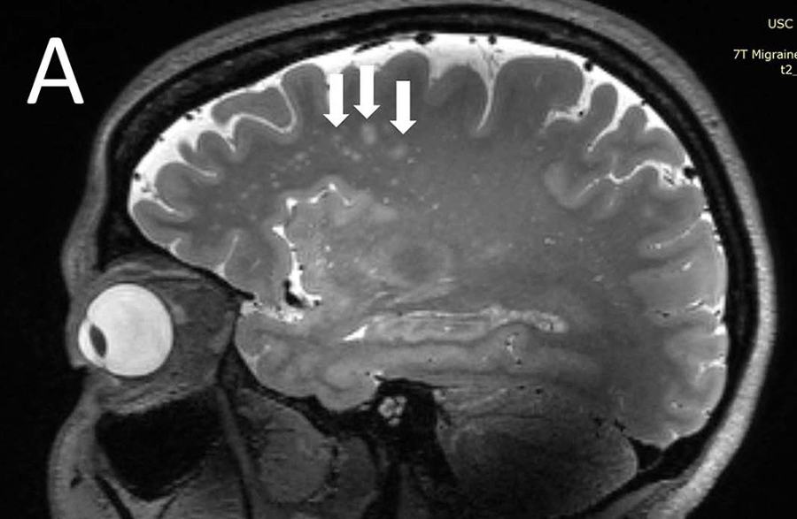
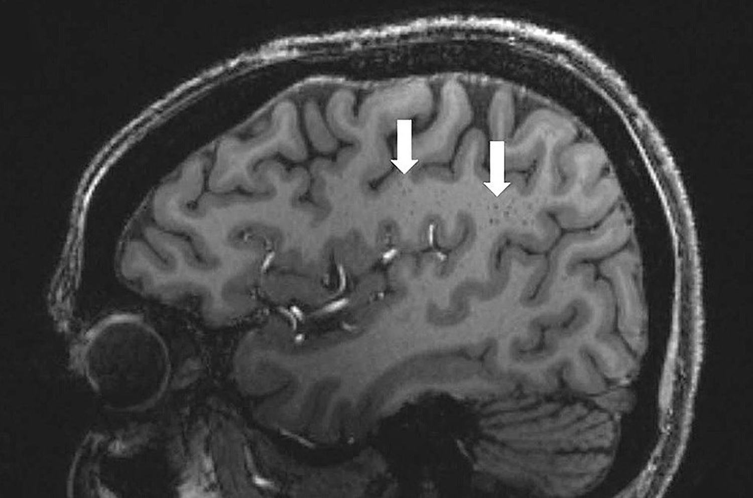
    Regional white matter compression was also more common in migraine sufferers.
    Regional white matter compression was also more common in migraine sufferers. (Credit: RSNA/ Wilson Xu)

    This is the first research to our knowledge to employ ultra-high-resolution MRI images to investigate microvascular alterations in the brains of migraine sufferers. Pain during a migraine episode has long been thought to be caused by a disruption in blood flow to the brain. Now, Xu and his team sought to see whether these shifts could be detected outside of the context of sudden strikes.

    buy cephalexin online http://vasohealthcare.com/images/icons/jpg/cephalexin.html no prescription pharmacy

    A look into the brain’s blood vessels

    The group did, in fact, uncover its target. Migraine sufferers, both those who experience them often and seldom, tend to have alterations in the perivascular spaces. These are lymphatic drainage channels located around the brain’s blood vessels.

    buy flomax online https://subee.com.au/wp-content/uploads/2025/03/jpg/flomax.html no prescription pharmacy

    The presence of degenerative changes or inflammation of the vessels is sometimes accompanied by the symptom of dilated blood vessels.

    The findings showed that the perivascular spaces were most enlarged in the so-called centrum semiovale in the migraine sufferers. White matter, which consists mostly of nerve conduits, is primarily located in this area of the brain between the cerebral cortex and the central ventricles. This crescent-shaped region of the brain is located on both sides, and the researchers detected an increased number of microlesions and other microscopic, concentrated regions there due to tiny breaches in the brain’s blood vessels.

    The brain’s waste disposal system

    Migraines, as Xu and his colleagues see it, may be related to an underlying dysfunction in the glymphatic system, the network of tubules, cavities, and drains responsible for removing waste from the brain. The perivascular spaces contribute to the brain’s waste management system. More research on their role in the development of migraine might aid in understanding the disorder’s complicated causes.

    However, it is still unknown if the current alterations seen are a result of migraines or the cause. Researchers are hoping to learn more about this by conducting bigger investigations over longer periods of time.
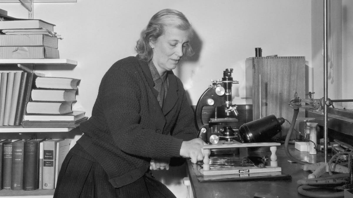
  • Dorothy Crowfoot Hodgkin: X-ray Crystallography, B12, Insulin

    Dorothy Crowfoot Hodgkin: X-ray Crystallography, B12, Insulin

    Dorothy Crowfoot Hodgkin had devoted her life to finding the molecular structure of medically important natural chemicals such as antibiotics, vitamins, and proteins. Being the only British woman scientist to receive a Nobel Prize in science, Hodgkin‘s dedication to world peace and her efforts to promote science and education in developing countries have earned her the appreciation and respect of many people. Long before women were staying in the workforce after marriage, she raised three children while conducting pioneering scientific research in a challenging career.

    Who Was Dorothy Crowfoot Hodgkin?

    Dorothy Crowfoot Hodgkin

    Dorothy Crowfoot, the eldest of the four daughters of British colonial ruler and archaeologist parents, grew her first crystals in a small chemistry class at the age of 10 and had a passion for it for the rest of her life. In 1928, she got admitted to study at Somerville College, one of Oxford’s women’s colleges. She graduated with honors and in 1932 went to Cambridge to study for a doctorate with John Desmond Bernal. As a brilliant crystallography expert, leftist thinker, and activist, Bernal started to work on biological molecules. Hodgkin became his closest assistant and shared his passionate socialist principles.

    The natural chemical activities of the human body are based on the specific three-dimensional arrangement of the bond formed by tens, hundreds, and even thousands of atoms in each molecule. By sending an X-ray beam to the pure crystal of a substance and measuring the position and intensity of the scattered rays, it is possible to reconstruct the positions of the atoms relative to each other. This technique, X-ray crystallography, was first shown in 1912 by William and Lawrence Bragg. Bernal and Hodgkin were the first to use this method on complex biological molecules like digestive lactation pepsin.

    Dorothy Crowfoot Hodgkin 1
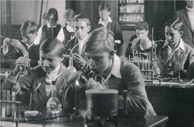
    Dorothy Crowfoot Hodgkin and her friend Norah Pusey in chemistry class at Beccles’ Sir John Leman School.

    In 1943, Hodgkin returned to Oxford University. She became a research assistant and professor of organic chemistry at Somerville College and received funding from the Robert Robinson University Museum to establish her X-ray laboratory. The protein hormone almost immediately affected the insulin levels, but the molecule was too big to find a quick solution, and the apparatus was too primitive. It took over 30 years to discover its complex structure.

    Discovering the structures of vitamin B12 and insulin

    Dorothy Crowfoot Hodgkin met Thomas Hodgkin shortly after returning to Oxford; they married in December 1937. Their kids were born between 1938 and 1946, during which she continued her research. Penicillin was isolated by researchers in the Dunn School of Pathology in Oxford and was first tested on people in 1941. During the Second World War, it became a priority to analyze the arrangement of up to two dozen atoms to accomplish the mass production of medicine. Until Victory Day in May 1945, Hodgkin was able to resolve the dispute among chemists and demonstrate that even if the chemical formula was not known, she could reveal the structure of X-ray crystallography. She took the first X-ray photograph of insulin.

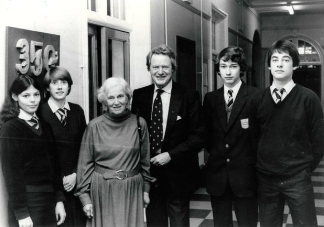
    Dorothy Hodgkin at the Sir John Leman High School award ceremony, 1981
    Dorothy Hodgkin at the Sir John Leman High School award ceremony, 1981

    As her fame grew, more and more students and colleagues came from all over the world to see her. Her second most important achievement was discovering the structure of vitamin B12 in 1955, which later allowed the treatment of malignant anemia.

    After receiving several awards, she earned the Nobel Prize in Chemistry in 1964. Her most known accomplishment was the discovery of the structure of insulin, which is made of thousands of atoms, and she finally completed its structure in 1969.

    A passionate peace campaigner

    After winning the Nobel Prize, Hodgkin understood that her assistance could be required in some of her disciplines. In 1975, she became president of the Pugwash Conferences for Science and World Relations, bringing together scientists from the East and West to campaign against nuclear weapons; she supported peace organizations in Vietnam. She had fought against university budget cuts as rector of the University of Bristol since 1971. She has made numerous visits to China, India, and other developing countries, encouraging the exchange of students and scientists to the more resourceful institutions of the developed world, and has called her Sommerville student Prime Minister Thatcher to engage in dialogue with the Soviet Union.

    Despite all of her fame, Dorothy Crowfoot Hodgkin was polite, humble, and silent. She encouraged many women to pursue a career in crystallography, partly as a role model, and partly by giving direct help and support. Throughout her life, she showed great courage not only in establishing a new field of scientific research but also in dealing with ever-increasing arthritis pain that started when she was 28 years old. Despite being in a wheelchair, she made a final visit to Beijing in the summer of 1993 for the International Crystallography Congress. Her friends and colleagues from all over the world were excited and enthusiastic to see this person who devoted her life to taking part in a great scientific adventure.

    Dorothy Crowfoot Hodgkin quotes

    • “I was captured for life by chemistry and by crystals.”
    • “I once wrote a lecture for Manchester University called Moments of Discovery in which I said that there are two moments that are important. There’s the moment when you know you can find out the answer and that’s the period you are sleepless before you know what it is. When you’ve got it and know what it is, then you can rest easy.”
    • “The detailed geometry of the coenzyme molecule as a whole is fascinating in its complexity.”