Tag: cell

  • History of Cancer

    History of Cancer

    Before the Common Era

    Traces of Cancer in the Remains of Ancient People

    In 1990, during excavations of the Chiribaya tribe cemetery in Peru on the northern border of the Atacama Desert, American professor Arthur Aufderheide discovered the mummy of a young woman with osteosarcoma — a malignant bone tumor. These remains were well-preserved thanks to the local climate: clay extracted all the liquid from the body, and the desert wind dried the tissues. However, scientists do not always find remains of tumor tissues. Sometimes, they come across traces of the disease left in the body of an ancient person, such as small holes in the skull and shoulder bones — results of metastases from skin melanoma or breast cancer.

    Incurable “Lumps in the Breast” from the Edwin Smith Papyrus

    One of the earliest written records of the disease is the Edwin Smith Papyrus, named after the American archaeologist who bought the artifact in an Egyptian market in 1862. This ancient Egyptian manuscript, dated to around 1600 BCE, is likely an incomplete copy of an earlier medical treatise created in the 27th century BCE. Its authorship is attributed to the famous Imhotep — an architect and healer of the Old Kingdom period. The surviving document describes 48 cases of various injuries, with the forty-fifth section unexpectedly devoted to the consequences of a severe specific disease — presumably breast cancer:

    When you examine the swelling lumps in the breast and find that they have spread throughout the breast, and if you place your hands on the breast, you find no heat and the tissues feel cool, with no graininess, internal fluid, or liquid discharge, but they protrude when touched, then you can say of the patient: ‘Now I treat the growth of tissues… the enlarged lumps of the breast mean the presence of swellings in the breast, large, spreading, and firm, and touching them is like feeling a ball of bandages, or they may be compared to an unripe fruit, hard and cool to the touch…’.

    For each injury described in the papyrus, the author suggests different treatments: for example, poultices for wounds and balms for burns. However, the mysterious “swelling lumps in the breast” baffled the ancient physician — unable to find a cure for this ailment, the “Treatment” section dryly states: “None exists.”

    The Persian Queen Atossa and the Successful Surgical Treatment of Breast Cancer from Herodotus’ “Histories”

    Two millennia after the Imhotep Papyrus, a similar disease reappears in written sources. Herodotus, in his “Histories,” tells of the Persian queen Atossa, who suffered from a bleeding breast tumor. Some researchers believe she had inflammatory breast cancer. None of the remedies helped Atossa, so the Greek physician Democedes proposed surgically removing the malignant tumor. The queen agreed, and the operation ultimately saved her life. Thus, Atossa’s story can be considered the first recorded example of a successful mastectomy — a surgical procedure to remove the breast.

    Ancient Rome

    Black Bile
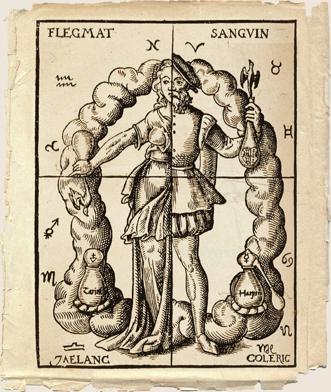
    The four humors. Illustration from Leonard Tourneisser's book Quintessence. 1570
    The four humors. Illustration from Leonard Tourneisser’s book Quintessence. 1570

    Roman physician Galen, who lived from 129 to 216 CE, following Hippocrates, believed that a healthy person’s body was in balance with four humors (fluids): blood, phlegm, yellow bile, and black bile. An excess of any humor inevitably led to illness, and treatment involved removing excess fluid from the body, for example, through bloodletting, emetics, and laxatives.

    Galen believed that malignant tumors were caused by an excess of black bile, which became a dense mass in the body. Interestingly, medieval physicians believed that an excess of black bile also caused melancholy: μελαγχολία in ancient Greek means “black bile.” Galen, whose authority remained unchallenged for over a thousand years, argued that the disease could not be defeated by surgery: the black bile would remain and pose a threat of new tumors. According to him, a patient could only be supported with general therapeutic measures.

    Middle Ages

    Boar Tusk, Fox Lungs, and Ground Elephant Bone

    Galen’s views predetermined the medieval medical approach to tumors. Surgery was generally considered more harmful than beneficial, and the absence of radical treatment was seen as the best possible remedy. An alternative to the surgeon’s knife was a wide range of rather exotic remedies: arsenic tincture, lead tincture, boar tusk, fox lungs, ground elephant bone, crushed white coral, castor bean seeds, senna plant, and more. Alcohol and opium tincture were used to relieve unbearable pain.
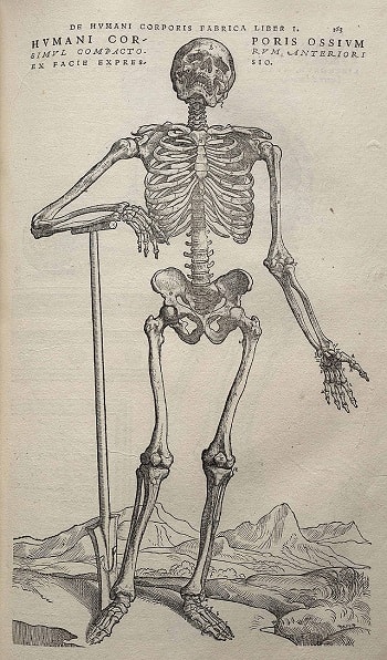
    Image from Andreas Vesalius' De humani corporis fabrica (1543), page 163
    Image from Andreas Vesalius’ De humani corporis fabrica (1543), page 163

    In the Renaissance, medicine revisited Galen’s idea that an excess of black bile in the body caused malignant tumors. Andreas Vesalius, the founder of scientific anatomy and author of “On the Fabric of the Human Body in Seven Books” (1543), who personally dissected corpses, could not find even the slightest trace of the infamous substance supposedly responsible for the development of tumors.

    Enlightenment: Cancer from an Excess of Lymph

    Another suspected cause of malignant tumors was lymph (also known as phlegm in humoral theory). According to this new understanding, tumors were no longer attributed to mysterious black bile but to a clear fluid that permeates the human body and is responsible for a calm temperament. German physicians of the 17th–18th centuries, Georg Ernst Stahl and Friedrich Hoffmann, suggested that tumors consisted of fermented lymph, which could have different densities, acidities, and alkalinities in each specific case.

    Cancer as Contagion

    While Hippocrates, Galen, Stahl, and Hoffmann sought the cause of the disease within the body, two 17th-century Dutch physicians, Zacutus Lusitanus and Nicolaas Tulp, found it outside. Independently, they reached the same conclusion: cancer is contagious. The doctors proposed moving all patients outside major cities to isolate them and thus prevent the spread of the dangerous disease. The idea of the contagiousness of cancer, prevalent in Europe during the 17th and 18th centuries, is now considered mistaken. However, it is known that certain viruses, bacteria, and parasites can indeed increase the risk of developing tumors, such as the human papillomavirus or the bacterium Helicobacter pylori.

    Cancer from Lifestyle

    In the 18th century, scientists made three important discoveries that laid the foundation for the epidemiology of cancer. In 1713, Italian physician Bernardino Ramazzini noted that cervical cancer was almost nonexistent among nuns, while the incidence of breast cancer was relatively high. Ramazzini concluded that lifestyle (in the case of nuns — lack of sexual relations) could directly influence the development of this disease in women.

    In 1775, English surgeon Percivall Pott discovered that scrotal cancer, often found in chimney sweeps, had an occupational nature: tumors arose due to the accumulation of soot in the folds of the scrotum. Pott’s discovery later led to the study of carcinogens associated with specific professions and the gradual adoption of occupational health and safety measures.

    By the 20th century, it was established that carcinogens could include benzene, used in the production of medicines, plastics, and rubber, as well as the fine-fiber mineral asbestos, used in construction, among others.

    It also became clear in the 18th century that bad habits could cause this disease. In 1761, English scientist John Hill, in his book “Cautions against the Immoderate Use of Snuff…,” first directly linked tobacco use and the development of tumors. However, lung cancer caused by smoking only became a subject of intensive research in the 20th century.

    Surgery as the Only Way to Defeat Cancer

    The medicine of the Enlightenment era drew a final line under Galen’s teachings. While Vesalius created a scheme of the “healthy” human body, the English anatomist Matthew Baillie managed to describe the “pathological” body in minute detail. In his work “The Morbid Anatomy of Some of the Most Important Parts of the Human Body” (1793), Baillie thoroughly examined various malignant formations, but in none of them could he find any hint of black bile.

    Having “buried” black bile as a scientific error, medicine paved the way for surgery as possibly the only effective method of fighting malignant tumors. However, as before, patients could only rely on the skill of doctors. Indeed, in the absence of means to alleviate their condition during operations, it was difficult to count on a favorable outcome: people who agreed to go under the surgeon’s knife typically died from pain shock, heavy blood loss, and various infections.

    19th Century

    Ether and Carbolic Acid

    The situation changed dramatically in the middle to late 19th century with the invention of anesthesia and antiseptics. American dentist William Morton began using the organic substance ether as a general anesthetic, while English surgeon Joseph Lister started using carbolic acid to disinfect wounds. After these important discoveries, doctors were able to resort to radical surgery when it was necessary to remove a tumor along with lymph nodes.

    Surgeons such as Theodor Billroth and William Stewart Halsted became famous in this field. Billroth performed the first esophagectomy (removal of part of the esophagus), laryngectomy (removal of the larynx), and gastrectomy (removal of the stomach) in history, while Halsted performed radical mastectomy, which is the complete removal of breast tissue.

    Cell Theory
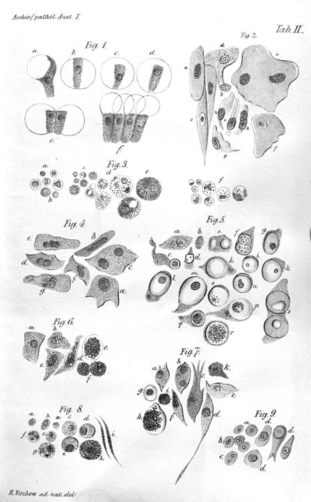
    Illustration of Virchow's cell theory
    Illustration of Virchow’s cell theory

    In the 19th century, as microscope design was improved, scientists began to gradually approach an understanding of the true nature of cancer. In 1838, German biologist Johannes Peter Müller, in his work “On the Finer Structure and Form of Morbid Tumors,” proved that malignant formations consist not of lymph but of cells. However, he still believed that tumor cells were formed not from normal cells but from blastema between them. But his student Rudolf Virchow proved that all cells, including tumor cells, are formed from other similar cells: omnis cellula e cellula (“all cells come from cells”).

    However, even Virchow was mistaken: he was sure that cancer spread through the body like a fluid. In the 1860s, German surgeon Karl Thiersch disproved Virchow, proving that tumors consist of epithelial tissue, not connective tissue. He showed that metastases appear as a result of the spread of malignant cells, not through some unknown fluid.

    20th Century

    Hormone Therapy

    In the late 1870s, English physician George Thomas Beatson discovered a direct connection between the ovaries and milk production in the breast: after removing this organ in rabbits, he noticed that they stopped producing milk. This discovery led Beatson to wonder: could oophorectomy, or removal of the ovaries, have a positive effect on advanced breast cancer? Indeed, his experiments showed that such an operation often helped improve the condition of women with this type of cancer.

    The scientist also suggested that the ovaries themselves might be the main cause of breast cancer development. Even before estrogen was discovered, Beatson determined that the female hormone from the ovaries had a stimulating effect on breast cancer. Beatson’s discoveries laid the foundation for modern hormone therapy, where substances like tamoxifen, which suppresses the effects of estrogen, and aromatase inhibitors, which inhibit enzymes that convert male hormones (androgens) into female hormones (estrogens), are used to treat and prevent breast cancer.

    In the 20th century, a hormonal method was also found to treat a male disease—prostate cancer. In the 1940s, American scientist Charles Huggins discovered that after castration, patients experienced a sharp regression of metastatic prostate cancer. In addition, it was found that simultaneously decreasing testosterone levels and increasing estrogen levels helps in treating “male” cancer.

    Radiation Therapy
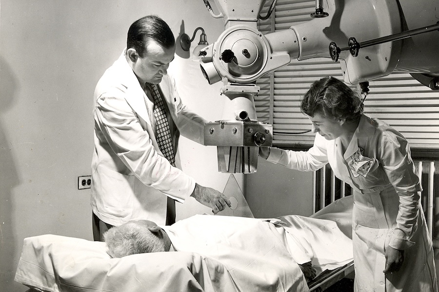
    Radiation therapy at MSK circa 1949
    Radiation therapy at MSK circa 1949. Image: Memorial Sloan Kettering Cancer Center

    At the end of the 19th century, Wilhelm Röntgen discovered X-rays, and Marie Curie and Pierre Curie discovered radium. Along with this came a new direction in tumor treatment: radiation therapy. Scientists found that radium could damage tumor cells in the body to the point of their complete destruction. However, in the early stages of radiation therapy, many doctors did not fully realize that radiation attacks healthy cells just as successfully as diseased ones. If the necessary dose is miscalculated, the rays can be deadly.

    It took almost a century to fully control unmanageable radiation. At the end of the 20th century, conformal radiation therapy was invented, in which the beam is precisely directed at tumor tissues thanks to detailed three-dimensional models created using computed tomography.

    Chemotherapy

    The militarism of World War II killed millions of human lives but unexpectedly helped find a new means in the fight against malignant tumors. In the 1940s, as part of developing more effective weapons, American scientists Louis Goodman and Alfred Gilman, commissioned by the US Department of Defense, studied chemical substances related to mustard gas, a poisonous chemical compound. During these studies, they accidentally found that nitrogen mustard helps in treating cancer of the lymph nodes (lymphoma). This was one of the first steps towards introducing chemotherapy into medical practice.
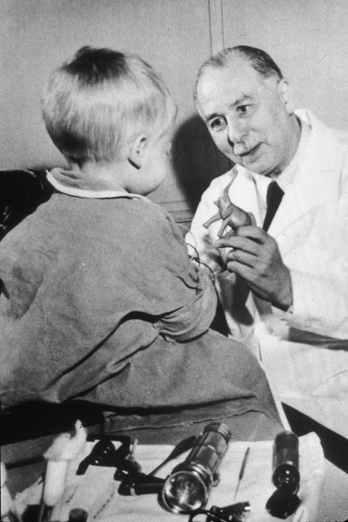
    Sidney Farber. 1960
    Sidney Farber, 1960. Image: Public Domain

    The real era of chemotherapy began with Sidney Farber, an American oncologist who, in the late 1940s, proved that a substance called aminopterin could cause remission in children suffering from acute leukemia: it blocks the division of leukocytes. Subsequently, adjuvant therapy became widely used in medical practice, which is a special chemotherapy aimed at destroying all tumor cells remaining in the body after surgery. Such therapy was first tested in the treatment of breast cancer, then in the treatment of colon cancer, testicular cancer, and other diseases. Another important innovation was combination chemotherapy, in which several different drugs are used simultaneously for more effective treatment.

    Immunotherapy and Targeted Drugs

    One of the most effective modern means in the fight against malignant tumors is immunotherapy. It has been actively used since the 1970s. Immunotherapy involves introducing special biological agents into the patient’s body that can both mimic the natural immune response and help the body’s own immune cells fight the tumor. The first targeted drugs—rituximab and trastuzumab—were created in the late 1990s; since then, they have been used to treat lymphoma and breast cancer, respectively.

    Lymph node with mantle cell lymphoma
    Lymph node with mantle cell lymphoma. Image: Gabriel Caponetti, CC BY-SA 3.0

    A promising direction in modern immunotherapy is the development of special cancer vaccines. For example, in 2010, the first vaccine was approved in the USA for a form of prostate cancer that no longer responds to hormone treatment. This drug, unfortunately, cannot kill the disease, but it helps the immune system fight tumor cells, prolonging the life of patients who seem to have a fatal diagnosis.

    Another important direction in modern immuno-oncology is the development of checkpoint inhibitors that regulate mechanisms that block the body’s immune response. The first of these drugs, ipilimumab, was approved in the USA in 2011. Now patients with diseases that were previously considered incurable have a chance for effective therapy. For example, former US President Jimmy Carter was able to get rid of metastatic melanoma when he was already over 90 years old.

    Also, quite recently, cell therapy using lymphocytes taken from the patient, which are then genetically modified, began to spread. It was first and successfully used against acute lymphoblastic leukemia in a five-year-old American girl, Emily Whitehead. In addition, research in the field of gene therapy continues. In general, modern medicine strives for ever-greater personalization of treatment, which makes it possible to completely cure the disease.

  • How pioneer factors dissolve the compressed genetic material

    How pioneer factors dissolve the compressed genetic material

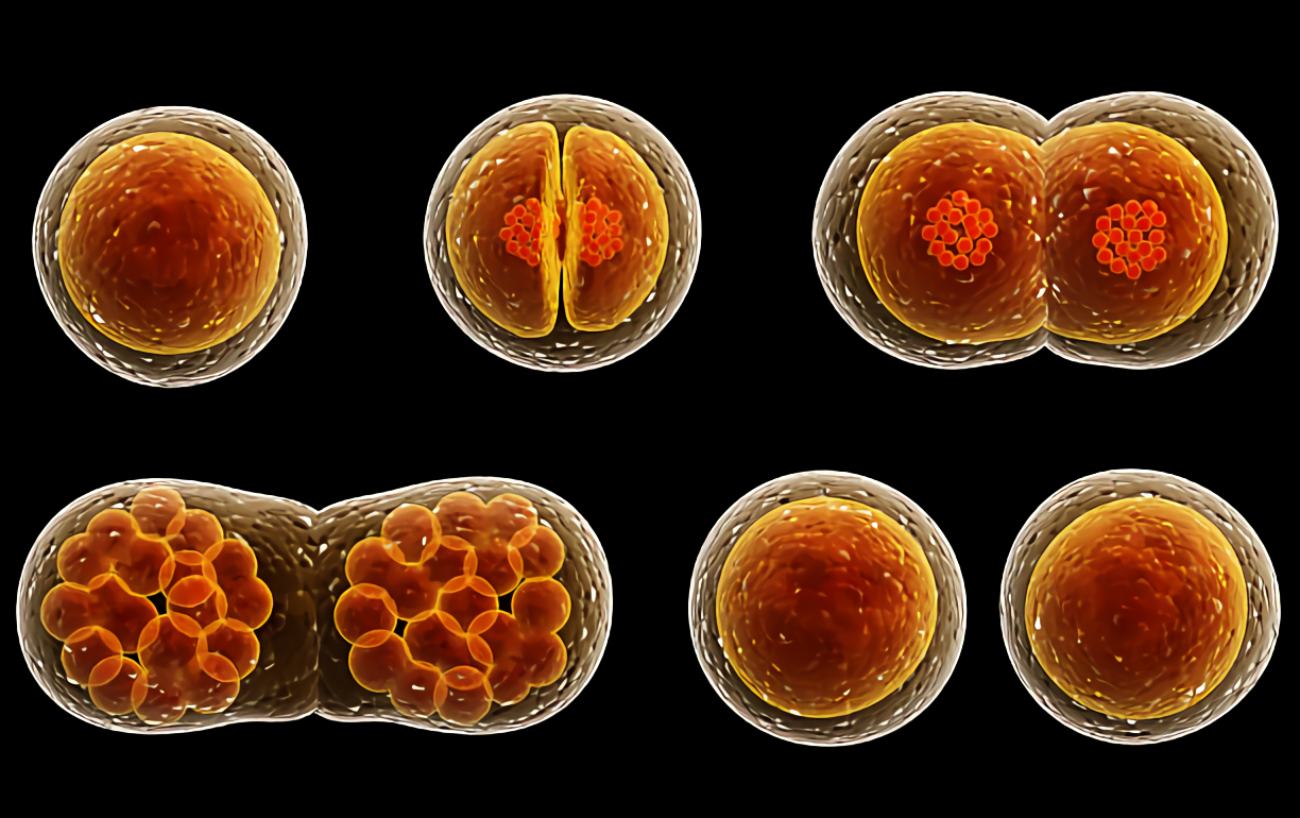
    A fertilized egg cell must decompress its genes before it can develop into an embryo. Scientists have finally figured out how it accomplishes this feat. According to the findings, a so-called “pioneer factor” is responsible for opening the chromatin packing of the genetic material during fertilization, allowing the genes crucial for early cell division to be read.

    buy bupropion online http://mediaidinc.com/scripts/js/bupropion.html no prescription pharmacy

    This is the only way the embryo can develop beyond the two-cell stage.

    If everything goes well, a new life starts when a sperm fertilizes an egg. Things may go wrong even when the mother’s and father’s chromosomes fuse together. However, the following phases are not without their own challenges: The newly fused DNA in a fertilized egg cell has to be “awakened” before it can divide. So-called pioneer factors are responsible for reading the relevant parts of DNA during cell division.

    The future embryo undergoes its first round of cell divisions shortly after fertilization. On the other hand, this requires the reading of at least some genes.

    buy ozempic online http://mediaidinc.com/scripts/js/ozempic.html no prescription pharmacy

    Obtaining the genetic material from its sealed packaging

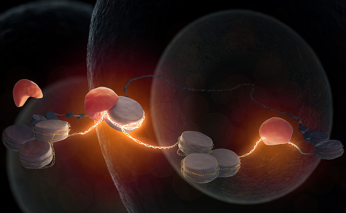
    In a fertilized egg cell, the pioneer factor Nr5a2 (red) interacts to the inactive DNA (gray) that is still wrapped around histones. Thereby, it wakes the genome. Essential genes for embryonic development may now be obtained.
    In a fertilized egg cell, the pioneer factor Nr5a2 (red) interacts to the inactive DNA (gray) that is still wrapped around histones. Thereby, it wakes the genome. Essential genes for embryonic development may now be obtained. (Credit: Max Iglesias)

    Molecules in the cytoplasm of the egg cells that are passed down from the mother are useful even during the first cell division. When an embryo reaches the two-cell stage, however, the cells are on their own. The thing is, the genetic code is not openly available in the nucleus of the cell. DNA is present as a long strand that is wrapped like a string of pearls around smaller packing proteins called histones, as explained by co-author and Max Planck Institute researcher Siwat Ruangroengkulrith.

    The embryonic cells need to decode the first genes from this packing at certain locations before they can make the transcription factors required for further cell divisions. But the DNA thread is shortened by as much as 40,000-fold when twisted into histones.

    buy zydena online https://mexicanpharmacyonlinerx.net/buy-zydena.html no prescription pharmacy

    It was previously unclear which unpacker molecules, in the form of so-called pioneer factors, liberate the compressed genetic information and allow for successive cell divisions.

    The first genes in a fertilized cell are read by a specific pioneer factor.

    The key molecule

    First author Johanna Gassler of the MPI of Biochemistry and her colleagues have shed light on this issue. First, scientists determined in mice which transcription factors are active throughout the development of an embryo and which genes were available for reading. Researchers sought for sequence features shared by the earliest-stage mRNA molecules they isolated and found many.

    A closer look at the molecules involved has highlighted the crucial function played by one in particular: Nr5a2, the pioneering factor. Embryonic development cannot go through the two-cell stage without it, since it is essential for activating the genome at that time. This pioneering factor seems to be transmitted from the maternal egg cell to the developing embryo.

    The absence of Nr5a2

    Without Nr5a2, no additional embryonic processes may begin. Through their tests, they demonstrated that inhibiting Nr5a2 prevents the production of the vast majority of mRNA molecules in early embryos. Embryos are also prevented from developing further. This indicates the significance of Nr5a2 in the first stages of embryonic development.

    The team was also successful in elucidating the mechanism behind this pioneering factor. Experiments have demonstrated that Nr5a2 may unlock dormant DNA sequences, allowing other genes’ transcription machinery to reach those regions. To begin the process of unpacking and reading the genetic information in embryos, the molecule docks onto the chromatin envelope of the DNA.

    buy mounjaro online http://mediaidinc.com/scripts/js/mounjaro.html no prescription pharmacy

    Additional transcription factors are then released, which control further reading.

    Relevant understanding of the first moments of existence

    Important progress has been made toward a mechanistic understanding of the origin of life with the revelation that Nr5a2 plays a vital role in genome awakening. Researchers believe there must be more components at play, but they haven’t been able to pin them down yet.

  • DNA: Genetic instruction manual of all organisms

    DNA: Genetic instruction manual of all organisms

    The written version of our genetic instruction manual, which has 3 billion letters, would take up many volumes’ worth of space in real life. However, it is only contained inside the tiny cellular structure of our body. From our gender to our physical characteristics to our susceptibility to disease, practically every aspect of our lives is determined by the choices our ancestors made. However, for a very long time, no one knew what this genetic code looked like and what it contained. Scientists eventually uncovered the shape, language, and exact function of our DNA, with some unexpected findings along the way.

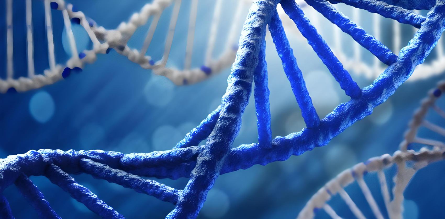
    The genetic specifications for all known creatures and many viruses are stored in a deoxyribonucleic acid or DNA, a polymer made up of two polynucleotide chains that coil around each other to create a double helix. DNA governs development, functioning, growth and reproduction.

    Two men did change the world of science

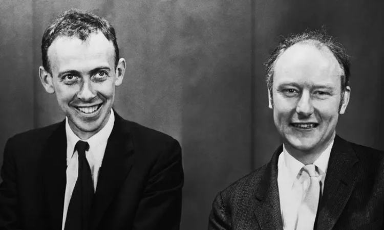
    james watson francis crick 2

    In a statement made 70 years ago, according to James Watson, only a few discoveries have been of such exquisite beauty as DNA. Watson was referring to the double helix, a structure that is 2.5 nanometers in diameter, looks like a helically twisted rope ladder, and stands 7,2 feet (2,2 meters) in length if fully unfolded. 

    On April 25, 1953, James Watson and Francis Crick published a single page in Nature proposing a model for the three-dimensional structure of deoxyribonucleic acid (DNA), the molecule that encodes human genes.

    It seemed that the two researchers were confident in the long-term relevance of their model since they cited “novel features of considerable biological interest” at the start of their paper.

    Zero interest in chemistry

    Although at first glance, it did not seem likely that two “scientific clowns,” as scientist Erwin Chargaff dubbed them, would produce such a groundbreaking discovery. James Watson, who was very talented, began studying biology at the University of Chicago when he was only 15 years old. Birds were his major focus at the time, thus he was able to avoid taking any science classes.

    The zoologist’s understanding of chemistry and physics was quite limited when he first arrived at the Cavendish Laboratory in Cambridge, England, in the autumn of 1951, at the tender age of 23. In England, he met British scientist Francis Crick, who was 13 years older and whose loud laughing was the bane of his colleagues’ existence. Francis Crick’s prior life as a researcher was summed up by the institute’s director, Sir Lawrence Bragg. According to him, Francis was talking ceaselessly and had come up with next to nothing of decisive importance.

    A scientific footrace

    In 1949, Erwin Chargaff discovered that the DNA bases adenine, thymine, cytosine, and guanine always occur in DNA at a 1:1 ratio, or most likely in pairs. The next step was to figure out the structural integrity of the bases and how they fit together. Watson, who was originally uninterested, attended a presentation by neighboring King’s College London scientist Rosalind Franklin in November 1951, during which she shared recent X-ray diffraction photographs of DNA.

    Waston found intriguing her speculation that DNA could exist in a twisty helical shape with two, three, or four twists. As soon as Watson and Crick got back to Cambridge, they set out to try to replicate this structure. They hypothesized, based on chemical calculations, that the structure would consist of three chains joined in a helix by magnesium ions, with the molecular arms pointing in all directions.

    Success through failure

    Watson, however, was not paying close attention, and the team’s model of the chemical reaction turned out to be incorrect. They made a disappointing appearance in front of Rosalind Franklin and London-based biophysicist Maurice Wilkins.

    Colleagues were quite harsh in their criticism. Previous X-ray images produced by these two scientists had demonstrated conclusively that the supporting chains could not lay within, refuting the premise of Watson and Crick, and that magnesium ions were scarcely capable of maintaining this structure.

    In July of 1952, Erwin Chargaff visited Watson and Crick in the lab and delivered a similarly damning assessment of their scientific prowess: “enormous ambition and aggressiveness, coupled with an almost complete ignorance of, and a contempt for, chemistry…”

    When it became public that famous scientist Linus Pauling, on the other side of the Atlantic, shared a fascination with the structure of genetic information and suggested a model for it, the scientific reaction intensified. Urgency necessitated swift action.

    Then, towards the end of 1952, Maurice Wilkins offered Watson and Crick an X-ray structural study of his colleague, Rosalind Franklin, which proved to be a pivotal event that ultimately led to triumph. It was, in fact, a picture of a recently discovered DNA structure. But this was all without Franklin’s consent.

    By the end of the study, the two scientists had reached a consensus: DNA is made up of two strands that intertwine with each other like rungs on a rope ladder. Hydrogen bonds hold their molecular appendages, the complementary bases, together. Finally, Watson and Crick assembled their metal double helix structure like pieces of a jigsaw. This variation won over even the most skeptical people.

    Recognition and respect

    Many scientists date the beginning of molecular genetics to the publication of the “Watson-Crick Model” of the structure of DNA. James Watson, Francis Crick, and Maurice Wilkins all split the 1962 Nobel Prize in Medicine and Physiology equally. In contrast, Rosalind Franklin, whose research offered the last vital piece of the puzzle, came up empty. Sadly, she passed away from uterine cancer in 1958, when she was only 37 years old, without seeing the fruits of her labor. It’s safe to say that most people nowadays have forgotten who Franklin and Wilkins were. But the names Watson and Crick will forever be linked to the double helix model of DNA.

    The exchange of information 

    Translation of genetic code

    Since 1953, scientists have understood the fundamental nature of our DNA and that it includes the blueprints for every aspect of our identities, from physical appearance to health. It was quickly understood that each base pair represented a different letter in the manual. The question is how to give form to these inscrutable directives and create a live, breathing, and authentic human being.

    In every human cell, two sets of 23 chromosomes are created when an egg from the mother and a sperm from the father fuse. The maternal contribution to these roughly X-shaped structures is half, whereas the paternal contribution is half.

    We store and carry our DNA, or genetic information, in a compact form called chromosomes. All of our DNA, together with its protective envelope structures, is stored in a very condensed form on the many chromosomes in our bodies.

    The sequence of bases as the alphabet of life

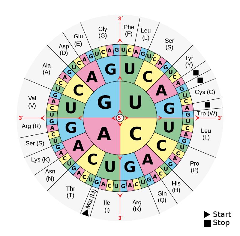
    Aminoacids.svg
    The typical RNA codon table is structured in the form of a wheel.

    Adenine (A), Guanine (G), Thymine (T), and Cytosine (C) are the four bases that make up DNA. The two strands of DNA’s framework are held together by the pairs A-T and C-G. They serve as the rungs on this hereditary ladder. When you align the ladder segments of a single DNA strand, you’ll see a lengthy string of base letters.

    And it is in them where the genetic code is found. Three of these letters are put together to spell out a word that specifies where in the process the production of protein should include a certain amino acid. A string of these letters forms a phrase, which in turn becomes the blueprint for a protein. And these molecules, in turn, play the role of a biochemical housekeeper who makes sure everything from cell and tissue formation to signal transduction and metabolic processes go smoothly.

    Transcription and translation of DNA

    Genes and proteins

    How, however, does the blueprint for a structure end up as a protein? The process of making proteins from scratch is called biosynthesis. At this phase, the genome must be unpacked so that the information for a protein can be read from DNA. Generally found as a double strand, DNA separates into two single strands. Thus, the free arms of the rope ladder become accessible.

    Beginning with the copy

    dna arm
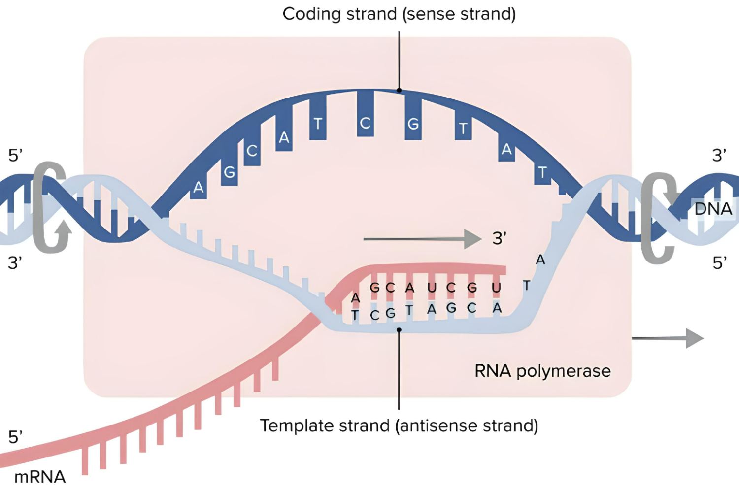
    Transcription produces messenger RNA. (Image credit: Lecturio)

    Enzymes have now made it possible to make carbon copies of this segment of the strand by simply joining a complementary base to each of the free arms. This time around, though, the base uracil bonds to the adenine instead of the thymine. In this case, however, the ribonucleic acid (RNA) serves as the scaffolding for these newly joined bases. After the copy is complete, enzymes cut the RNA strand and its accompanying DNA bases away, creating a copy of this region of the genome that can be moved throughout the cell, the messenger RNA (mRNA). Transcription refers to this process of making a copy of the genetic material and rewriting it.

    Translation into protein building blocks

    Ribosome mRNA translation
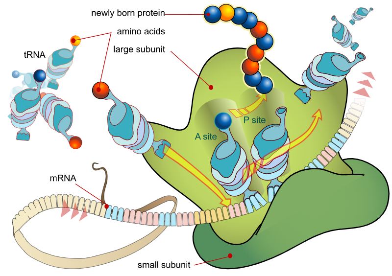
    A ribosome’s translation of mRNA and protein synthesis is shown in this diagram.

    However, this is just the beginning. mRNA now transports the genetic information copy from the nucleus into the cell plasma, where it will be read by the ribosomes and used to make proteins.

    The mRNA is sandwiched between the two subunits, one larger than the other; this creates a reading unit similar to the needle on a tape recorder. It decodes the genetic code by identifying which of the three bases (and hence which genetic code word) is present in each instance.

    Simultaneously, many of the amino acids that will make up the future protein accumulate on the ribosomes, each of which has a tiny piece of RNA consisting of precisely three base letters connected to it. These letters and numbers serve as a label, identifying the specific amino acid bound to this transport RNA (tRNA). It is the job of the ribosome to dock and connect the component of the amino acid that corresponds to the coding of the next amino acid in the read-out mRNA.

    Polypeptides, or chains of amino acids, are produced in this fashion and are the building blocks from which proteins are assembled. DNA can only perform its job via translation, the process by which the genetic information is converted into a chain of amino acids.

    Junk DNA

    Discarded material transformed into a control center

    A gene is a set of instructions for making a particular protein; it consists of a specific sequence of the base pairs cytosine, adenine, guanine, and thymine. It’s the blueprint for these critical messengers of our body’s processes.

    It was quickly discovered, however, that significant portions of human DNA lacked any recognizable construction instructions. Sequences in an organism’s DNA that do not code for proteins are known as noncoding DNA (ncDNA). They looked to be made up of illogical and repeated DNA sequences that had no discernible purpose. Therefore, scientists called these pieces of DNA “junk DNA.”

    Only 2% of your genes are real

    However, scientists were baffled when they looked more closely at the breakdown of our genetic material and saw that around 44% of it is “junk” in the form of several copies of genes and gene fragments (repeats).

    In addition, 52% seems to be useless as well and does not code for proteins. However, only around 2%–4% of human DNA is made up of genuine protein-coding genes.

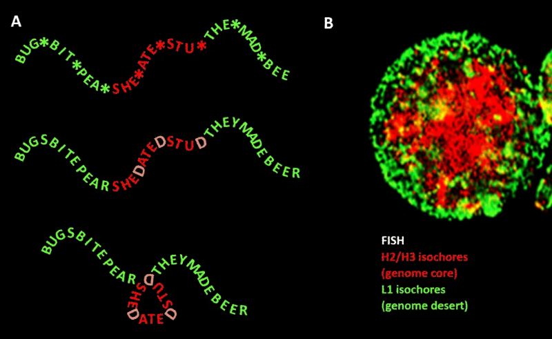
    It has long been a puzzle as to why evolution has preserved so much irrelevant DNA in addition to these gene sequences. But this issue was first answered by research in 2004. Scientists in the United States revealed a startling discovery about this “living genome deserts” that many regions of DNA that do not code for proteins were far from inactive. They include sequences that may activate or silence other genes, even if they are located far away.

    A regulator made of “junk”?

    This suggests that the genome’s so-called “junk” is playing a significant role in regulating gene activity, helping to shed light on the basic differences across species even though their genes are, on average, just a few percent different.

    Also, scientists from LLNL and JGI found that different parts of junk DNA have experienced different degrees of modification during evolution. There are several non-coding regulatory elements in the “desert areas” that are resistant to rearrangement and defend themselves via repeating junk DNA patterns. It appears that genomic regions known as stable genome deserts are essentially hidden gene regulatory components that preserve the intricate function of neighboring genes.

    About two-thirds of the genome deserts and about 20% of the overall genome could be gene segments that are completely useless for biology, indicating that most of the genome is redundant. At least, 75 percent or more of our genetic material is really just junk and only around 8–14% of our DNA is functional in some way.

    Our genome is governed by junk DNA

    junk dna
    Junk DNA and genome desert.

    The term “junk DNA” refers to the 98% of the human genome that does not code for proteins but the truth is actually more complicated. 

    Because this notion of mostly useless junk DNA kind of shattered in 2011. The international ENCODE project discovered something astounding: almost all of our junk DNA functions as a massive control panel for our genome, containing millions of molecular switches that can activate and deactivate our genes as needed, including in regions where only an “unstable desert” had been suspected.

    The “junk” has millions of switches

    Scientists created a detailed map of the locations and distributions of control elements, which revealed that the control switches are often located in inconveniently distant genomic regions from the genes they regulate. However, due to the complex three-dimensional shape of the DNA strands, they can still come close and exert their regulatory effects.

    That means, our genome is only functional because of switches: millions of buttons that control which genes are active.

    Genes derived from junk DNA

    But junk DNA has other, non-regulatory functions too; scientists from Europe identified a gene on mouse chromosome 10 that appeared out of nowhere but originated between 2.5 and 3.5 million years ago via genome-wide comparisons. The gene was the only one positioned in the center of a lengthy non-coding chromosomal part. This area is present in all other mammalian genomes as well. However, the gene is only found in mice.

    There was some speculation that a gene may emerge at a place in the genome that had never been used before, but no evidence for this had ever been found. Yet it was discovered that the mutations that only occur in mice could be responsible for the new formation of the gene.

    This demonstrates that the regions of DNA that do not code for proteins are an essential component of our genome and that they have long played a significant role in a variety of modern genetic analyses.

    DNA and forensic science

    It was all solved via a DNA analysis

    fingerprint

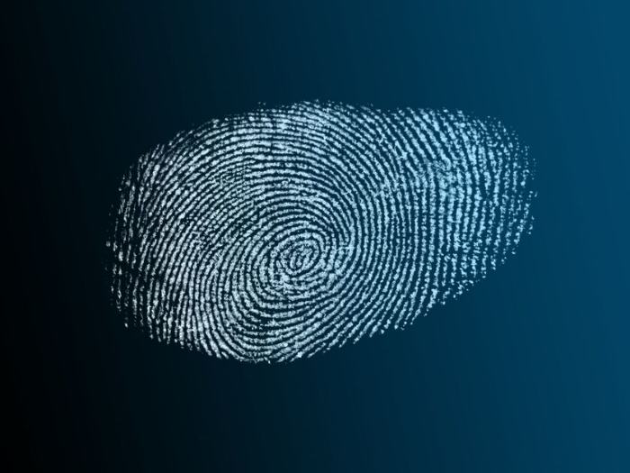
    You can identify a criminal by his genetic fingerprint from as little as a drop of saliva on a Coke bottle or cigarette filter, a few skin cells beneath the victim’s fingernails, or blood on his clothes.

    Most of our bodily fluids also contain cells from our body, and with them our genetic information. Skin cells are always left behind when a hand is dragged along a rough surface or is scratched, and these cells carry our DNA.

    However, there is a catch: there is far too little genetic material in the crime scene that remains for analysis, and this is precisely the reason why DNA analyses from such relics were unattainable for a long time. These genetic material remains can tell investigators whether or not their suspect was the perpetrator.

    The polymerase chain reaction for DNA testing

    But in 1983, US scientist Kary B. Mullis came up with a plan to multiply the few amounts of DNA that obtained and, in the process, devised one of the most pivotal techniques in genetics and biotechnology: the polymerase chain reaction (PCR).

    A DNA fragment of up to 3,000 base pairs in length is heated to 201 to 205 degrees Fahrenheit (94 to 96 degrees Celsius), which breaks the hydrogen bonds between the bases of the double strand, resulting in the separation of the helix into two single strands. Two primers are then added to the DNA solution.

    They bind to certain places on the DNA segments (based on their structures) and signal the beginning of the copying process, which is carried out by a heat-stable enzyme called polymerase.

    At a temperature of 140–160 degrees Fahrenheit (60–70 degrees Celsius), it joins DNA-building components floating in solution to produce a perfect replica of the sequence designated by the primers, leading to another double strand and doubling the original amount of sequences.

    Once the PCR is finished, the few remnants from the murder scene become a solution containing millions of copies of the perpetrator’s DNA, thanks to the process of repeated cycles in which the double strands are split from one another and then supplied with new halves by the polymerase.

    A unique repeat pattern

    The testing phase can now begin, with researchers comparing only small fragments of the DNA rather than the full sequence (which would take too long and be too laborious).

    These fragments are found in the genome’s non-coding regions and are made up of several repeating base sequences termed “short tandem repeats” (STRs), which provide a unique genetic fingerprint since their numbers vary from person to person.

    In many countries, a standard DNA analysis at a criminal lab includes testing for eight STR systems over several chromosomes and one sex-differentiating characteristic, which should be more than enough to rule out the possibility of a chance match.

    Estimates suggest that the number of people with whom our unique STR pattern is shared is less than one in a billion, with the exception of identical twins. If a suspect’s genetic fingerprint matches that found at the crime scene, then it’s likely that s/he committed the crime in question; her/his own DNA has in fact convicted him.

    Probing the paternity of a child

    The mother’s identity is generally evident since she gives birth to the kid (barring surrogate moms), but the identity of the father is not always so clear.

    It’s possible that the question of paternity won’t come up until the child is an adult if the woman has cheated on her partner in secret or if she gets pregnant shortly before breaking up with her partner and keeps the baby from him.

    Numerous laboratories around the world have long offered such gene-based paternity tests online, and the process for those willing to take the test is very simple: just send in a saliva sample, a few hairs with a hair root, a baby’s pacifier covered with spit, or a piece of chewing gum that has been well chewed.

    First, the DNA is extracted from the samples and amplified by polymerase chain reaction (PCR) in the lab; next, the DNA is compared to samples of the same genetic material from the child’s father or, ideally, the mother.

    Short tandem repeats (STRs) are also used in forensic DNA analysis, and the frequency with which a given base sequence is repeated within an STR marker varies from person to person but is passed down from parents to offspring. Each person carries two STR marker variants at each gene locus, one inherited from mother and one from father.

    In contrast, if the genetic material of the parent and child differs at three or more STR markers, paternity or maternity is considered to be ruled out. The probability that two unrelated people will have the exact same pattern of repeats at these markers is just one in 100 billion, according to current estimates.

    The Human Genome Project (1990-2003)

    The Human Genome Project 1990 2003 2
    Human Genome Project. Image credit: Encyclopædia Britannica, Inc.

    Learning by reading life’s book

    In the year 2000 in the United States, Bill Clinton and his British counterpart, Tony Blair, arranged for an unusual news conference in Washington. Nothing less than the human DNA itself was at stake here. The decoding of our genetic composition has been publicly announced by Clinton and, following him, by representatives of two rival research organizations, one government and one private.

    And in 2022, scientists finally announced that they finished decoding the entire human genome. According to that, about 30,000 human genes are housed in the nucleus of each human cell, where they are contained in 23 chromosomal groups.

    Humanity’s next big thing

    An early version of the “Book of Life” has been deciphered by both the worldwide Human Genome Project (HGP) scientists and genetic engineering pioneer Craig Venter and his business Celera. About 3.1 billion letters make up our genome, which is composed entirely of apparently random sequences of the four nucleotide bases (adenine, cytosine, guanine, and thymine).

    From the neurons that carry impulses throughout the brain to the immune cells that help protect us from external attack, each of the trillions of cells that make up our bodies has the same 3.1 billion DNA base pairs that make up the human genome.

    It’s still not fully known what words and sentences may be constructed from these letters, as well as where certain functional units of genetic material are buried.

    The decipherment of the human genome paved the way for novel approaches to illness prevention, diagnosis, and treatment. But these 3.1 billion letters of sequence in one human DNA were only the beginning of the long road to deciphering the human genome.

    Interesting, but impossible

    Things looked very different 25 years ago. In 1985, a group of genetics experts at the University of California, Santa Cruz, were approached by biologist Robert Sinsheimer with an unusual proposal: Why not try to sequence the human genome? The response was as unanimous as it was unequivocal: bold, exciting, but simply not feasible. Decoding even small sections of DNA was still too laborious at this time.

    However, one of the researchers involved, Walter Gilbert of Harvard University, did not give up on the idea. About 20 years ago, he and a colleague were the first to develop a method for reading out the genetic code or genetic sequencing.

    However, potential backers were still cautious, asking, “What if it turns out that the entire thing is not worth the massive effort?” and “Shouldn’t we possibly start with the genome of a small, less sophisticated creature, such as a bacterium?”

    Genome arms race

    Finally, in 1988, the U.S. National Institutes of Health (NIH) was convinced to organize a project to decode the human genome, led by none other than James Watson, one of the two discoverers of the double helix structure of DNA.

    Understanding of the disease genes

    However, progress has been sluggish since researchers were always debating whether or not it would be more efficient to begin by searching for illness genes rather than meticulously sequencing everything.

    Craig Venter, a geneticist at the National Institute of Neurological Disorders and Stroke (NINDS), stood out because he and his colleagues had created a novel approach to discover gene fragments at an unparalleled rate, but without understanding their function.

    Watson opposed and publicly complained about the sellout of genetic material which was met with early enthusiasm by NIH leaders since, if patented, these genes could be converted into cash. The fallout was seen when Watson was replaced as project head by Francis Collins in April 1992.

    Not fast enough

    Collins made a dismal prediction in 1993 that human genome sequencing wouldn’t be finished until 2005 at the earliest if things kept moving at their current rate. Part of the reason for this was the lack of resources that have so far prevented the development and widespread use of state-of-the-art DNA sequencers, which would greatly facilitate the automation of the genome decoding process.

    On the other side, achieving a success rate of 99.99 percent was a need. After all, international research institutes were joining the effort at an increasing rate.

    Upon meeting Craig Venter in 1995, the HGP researchers and management were rudely roused. As part of his new job at a commercial corporation, Venter released the first genome of a fully developed organism, that of the Haemophilus influenzae bacteria. He had accomplished it in a year because of the cutting-edge computing power available at the time. While progress was being made by Collins and the HGP researchers, it was slower than some would like.

    Head to head

    At the 1998 annual gathering of genetic experts, Venter pulled off his next move by announcing that his new firm would be able to decode the human genome in three years for a quarter of the cost of the HGP. He would be assisted by an automated sequencing system currently under development.

    At this point, Collins and his group must take action. Six months later, they announced that instead of waiting until 2005, full genome sequencing was now expected to be completed in 2003 thanks to increased efforts. They wanted to provide the first functional version of the human genome that is around 90% valid by spring 2001.

    It seemed like Craig Venter and his business, Celera, were in for a close finish. In reality, though, efforts to establish a mutually agreeable human genome resolution had already begun behind the scenes.

    The HGP suggested holding a combined press conference to announce the initial versions of both projects at the same time on July 26, 2000. While the HGP had been publishing their sequencing in the British journal “Nature,” Venter and his colleagues had been contributing to the rival American journal “Science.” The unveiling of the virtually entire human genome was announced two years ahead of schedule, on April 14, 2003.

    Thus, in April 2003, the Human Genome Project (HGP) was announced as completed but only around 85% of the genome was actually included. 15% of the remaining human genome was sequenced only by January 2022.

    Dictionary of genetics

    Amino acids

    20 amino acids are the fundamental building blocks of proteins, and the genes determine the order in which these amino acids are put together to create a chain.

    Bases

    The nucleotides adenine (A) and thymine (T) and cytosine (C) and guanine (G) are paired with one another in double-stranded DNA through the complementary base pairing concept. Thymine (T) is switched out for uracil (U) in the ssRNA (single-stranded RNA).

    Chromosomes

    Chromosomes, which contain an organism’s genetic information, number 46 in humans thanks to the duplication of the 23 chromosomes found in each of our cells.

    Codon

    An amino acid’s genetic code is encoded in a sequence of three bases.

    DNA

    Deoxyribonucleic acid is a double-stranded molecule composed of a sugar backbone (deoxyribose) and a phosphate group, and a linear series of base pairs. The two single strands are complementary to each other, run in an antiparallel direction, and are kept together by base pairs.

    DNA sequence

    An order of the DNA molecule’s construction order.

    Dolly

    A well-known cloned sheep that was cloned in 1996 from an adult sheep’s single cell.

    Gene

    Genes are sections of DNA. In eukaryotes, genes are often made up of coding sections (exons) and noncoding sections (introns). Coding portions (exons) carry the genetic information for creating proteins or functional RNA (e.g., tRNA).

    Genetics

    Molecular genetics investigates the fundamental laws of heredity at the molecular level, whereas classical genetics focuses on the inheritance of characteristics, especially in higher species. Applied genetics focuses on the breeding of economically highly productive crops and animals.

    Genetic code

    The genetic code is a kind of encryption used to store information on DNA, and it is represented by a set of three-base pairs in all known forms of life.

    Genetic fingerprint

    Genetic fingerprints are unique to each person and are generated by using so-called restriction enzymes and undergoing further analytical processes.

    Genome

    A genome refers to the whole set of genetic instructions for a certain organism.

    Human Genome Project

    An international effort funded by many agencies to investigate the DNA sequence, protein function, and regulatory mechanisms of the human genome.

    Gamete

    Gametes are sexually reproducing cells (eggs, sperm) that contain just one copy of each of the 23 genes found in the human genome, which is called haploid (in humans).

    Cloning

    Producing offspring with the same genetic material by cell division or nuclear transplantation.

    Mutation

    Mutations, which may be caused by anything from exposure to ultraviolet light or naturally occurring radioactivity to the simple passage of time, are the fundamental mechanism by which new species are created and evolve.

    Nucleic acids

    Both DNA and/or RNA

    Nucleotides

    A phosphate group, a sugar, and a base make up the three components of the DNA-building block.

    Nucleus

    The nucleus is the membrane-bound organelle that houses the cell’s chromosomes.

    Peptides

    Peptides are compounds made up of two or more amino acids, which can be the same or different. Peptides are classified according to their length, with dipeptides consisting of two amino acids, tripeptides of three, oligopeptides of two to nine or ten amino acids, polypeptides of ten to ninety-nine or one hundred amino acids, and macropeptides of one hundred amino acids or more being considered proteins.

    Polypeptide

    Chain of ten or more amino acids held together by peptide bonds.

    Polymerase

    The protein-making enzyme uses DNA as its template.

    PCR (Polymerase Chain Reaction)

    In 1985, Kary Mullis devised a method of enzymatically amplifying tiny amounts of DNA to provide enough material for genetic analysis of nucleic acid sequences.

    Protein biosynthesis

    Translation and transcription are two steps in the protein production process, which takes place on ribosomes inside a cell. Enzymes, hormones, and antibodies are all examples of proteins. Protein is a class of molecules that is predominantly made up of 20 distinct amino acids.

    Proteome

    Complete set of proteins in a cell, organ, or tissue fluid.

    Purine bases

    Adenine and guanine are two examples of purine bases.

    Pyrimidine bases

    DNA and RNA both use the pyrimidine nucleotide uracil, however, RNA uses cytosine and DNA uses mostly thymine.

    Restriction enzymes

    DNA scissor enzymes are enzymes that detect a particular sequence of letters on DNA and cut the DNA at that sequence.

    Ribonucleic acid (RNA)

    Ribonucleic acid (RNA) is the “little sister” of deoxyribonucleic acid (DNA), a single-stranded nucleic acid molecule involved in protein production in which the nucleotide uracil (U) replaces thymine.

    Ribosome

    The ribosome is the cell’s “protein factory,” where proteins are made by reading a copy of a gene.

    Telomeres

    Normal cells may undergo around 2,000 cell divisions before showing signs of wear and tear, during which time DNA ends (telomeres) that do not carry genetic information shrink.

    Transcription

    The overwriting of a gene’s DNA into messenger RNA (mRNA).

    Translation

    The method carried out by ribosomes whereby a protein is synthesized from its constituent amino acids.

    Virus

    Pathogenic biological structure made up of proteins and nucleic acids that may infect, replicate, and kill host cells.

    Viruses are dangerous because they rely on a “host” organism for their metabolism.

    Cell

    DNA is packed into chromosomes in the cell nucleus, making the cell the smallest reproducing unit in higher animals.

  • How Does Copper Kill Microbes Like Bacteria?

    How Does Copper Kill Microbes Like Bacteria?

    Copper has been a sought-after raw material for ages, and for good reason. It is used in a wide variety of products, from jewelry and coins to brass and electrical cables. However, the bright red precious metal has another use in medicine; it may be used as an antimicrobial agent. Pathogens such as bacteria are reportedly rendered harmless by the surface of the copper. But yet, could this really be true?

    Unquestionably, it is the case. An antibacterial effect can be seen in both copper and silver. Copper metals are effective against numerous types of bacteria, viruses, algae, and fungi. This is due to the several mechanisms through which copper toxicity affects these bacteria. Therefore, microorganisms have a hard time devising countermeasures to this kind of assault.

    How does copper kill microbes like bacteria?

    Copper Sulfate Rather Than Chlorine for the Pools

    Even though copper’s antimicrobial properties are not well understood, they have been known for quite some time. Even Roman legionaries understood that water stored in copper vessels would not rot on the journey back to camp. Honey and copper powder were a common antibacterial combination used by the ancient Greeks. Especially in the last two decades, copper and silver electrodes have been used by many swimming pools in place of chlorine to disinfect the water.

    In fact, a quick experiment can reveal whether or not copper is really effective as a natural antimicrobial. You can drop down a bacterial solution on a copper surface and let it dry. In just a few minutes, the number of bacteria cells in the drop will decrease by an order of magnitude or more. If there were 100,000 bacteria cells at the beginning, there wouldn’t be a single one left at the end.

    What Makes Copper So Harmful to Microorganisms?

    How do the antibacterial properties of copper work? It is the copper ions that make the difference. When the copper comes into touch with water, the copper ions are released. Bacteria is just a little water bag. The microorganisms are now subjected to a barrage of attacks from the freed copper ions. With enough force, they may breach the bacterial cell wall, allowing its contents to spill out.

    Copper is effective both externally and internally. Bacteria actually need a certain quantity of copper to function. Thus, they’ve evolved pumps in their cell wall to bring copper ions inside. And if there are a lot of ions in the environment, the microorganisms take in more copper than they should.

    As a result, copper kills off the microorganism’s intracellular proteins and even assaults DNA, the cell’s genetic material. Ultimately, this causes the bacteria to die.

    Copper as an Antimicrobial Building Material

    Copper offers several advantages over traditional antimicrobials, particularly in healthcare settings but also in the home or in public places. Alcohol and other antimicrobials are ineffective because they evaporate too rapidly to maintain their effectiveness for a long time. A copper surface, such as a door handle or light switch, on the other hand, is actually constantly killing germs.

    There is, however, a catch when it comes to such use cases. Copper surfaces degrade faster over time when exposed to sweaty hands and cleaning chemicals. A portion of their antibacterial function is lost when they form a dark coating with time.

    Scientists are trying to improve copper and copper alloys so that they retain the same level of antibacterial activity throughout time.