With only a sideways look, we know our hearts will soon be racing. It’s not just anyone’s gaze, but only the look of the one we love. But, what is the biological reasoning behind this phenomenon? Why does the heart start to beat faster if we are in love?
How sweet it is: The finding of a soul mate, whether it be love at first sight or just love, induces an involuntary increase in our heart rates. To the point when our hearts begin to race. Meeting a soulmate has actually a solid scientific foundation, despite its artistic overtones.
When Honoré de Balzac said, “Love has its own instinct, finding the way to the heart,” he wasn’t sure he was stating it correctly. But drenaline, a chemical widely known to those in search of excitement, is directed only to the heart.
There’s an adrenaline surge at play when seeing a loved one, which causes your heart rate to spike to a hundred beats per minute.
Sequences of physiological events
As soon as two people who are meant to be together lay eyes on one another, an irresistible process is set in motion inside their bodies. The adrenal glands, which are placed above the kidneys as their name suggests, receive the first impulses from the brain. Their internal organ, the adrenal gland, is the primary source of the hormone adrenaline, which plays a key role in the body’s ability to deal with stress.
Hormones including adrenaline, epinephrine, and norepinephrine are secreted by the adrenal gland in response to impulses from the brain. As they circulate throughout the body, they make the heart pump harder and quicker.
After being secreted into the circulation, the hormone eventually reaches the heart. Like an arrow from Cupid, it pierces the air and lands at the center of our being. The end result is an increase in heart rate that is similar to that achieved after a session of running, but with less positive health effects.
That’s why tender-hearted individuals should not be involved in such phenomena. People with cardiac conditions should avoid such a rapid heart rate because of the risks it poses. Those who have had a heart attack in the past should be cautious, since the opposite of ecstatic love might result from their romantic endeavors.
The good news is that the danger of a heart attack can be greatly reduced with only a few days of therapy with beta-blockers. Who would have believed that having sex immediately after a stroke may be beneficial?
The side effects
Crushing and love at first sight both have serious cardiovascular repercussions. OCD sufferers tend to have lower than average serotonin levels, and love has been shown to have this effect.
The reciprocal desire that develops between the two lovers can be explained by a decrease in levels of this neurotransmitter, which helps regulate mood. It’s possible that this is the reason why, in the early stages of a romantic relationship, there aren’t many other things that catch our attention outside of our partner. How sweet it is. It renders us blind but also utterly obsessed.
Multiple hundred muscles can be found in a single individual. They enable our body’s motion and the operation of vital organs like the heart. However, not every muscle is built the same. Which muscle do you think is the most powerful?
There are more than 600 muscles in the human body that we can’t directly see. They account for well over half of an adult’s entire body weight. And they play a significant part, too. Our ability to move is thanks to the contractile cells found throughout our bodies’ connective tissues. Additionally, they let the heart do its job.
However, not all muscles are created equal; in fact, there are distinct kinds of muscles that each have their own set of characteristics. Which muscle, therefore, is the strongest in the human body? The tongue is not the correct response. Although it may seem like a single muscle, the tongue is really made up of 17 different muscles and is thus classified as an organ.
The human body muscle that has the most strength
Gluteus maximus.
Some would think of muscles in the arms, such as the biceps or triceps. However, the most potent muscle is not up there at all; rather, it is buried deep inside our buttocks. Specifically, this refers to the largest muscle in your buttocks, also known as the gluteus maximus.
The pelvic girdle, the group of bones that run from the hips to the base of the spine, is where this muscle is found. Located in the leg’s dorsal and lateral muscles, it helps move the knee and ankle. It is the biggest muscle in the body and has the shape of a square. The buttocks’ round, protruding form is also largely due to it.
The function of the gluteus maximus
The gluteus maximus is a strong muscle for a reason; it serves an important function. Indeed, it helps to keep the posture upright by stabilizing the basin and keeping the trunk in extension. More importantly, it plays a role in gait and athletic performance by facilitating thigh motion during walking and running.
Gluteus maximus motion is also required for bending the knees and standing up. As a matter of fact, the muscle is used in a wide variety of sports and activities, such as jogging, cycling, skiing, and rugby. It seems logical that it would be the strongest.
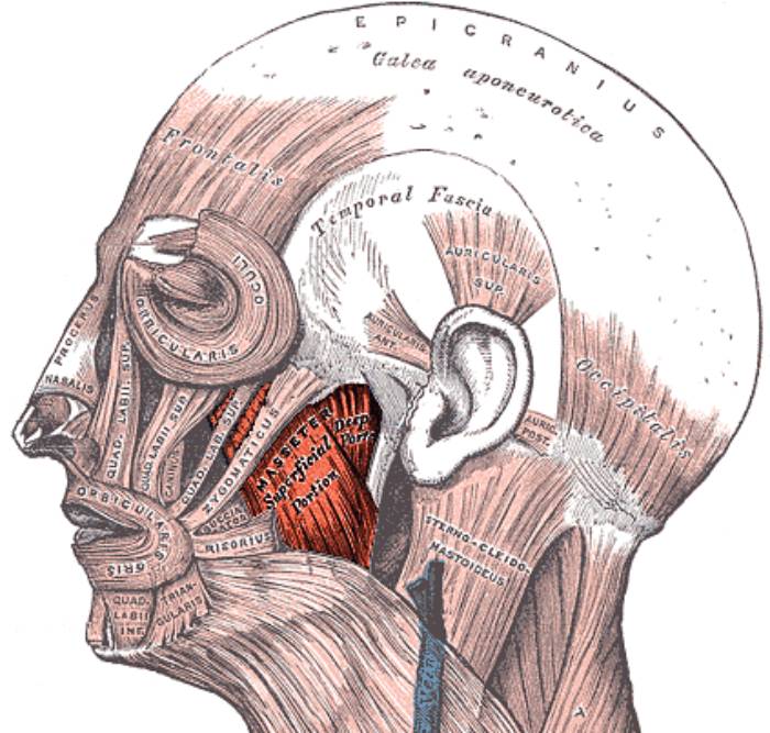
The masseter is the most powerful muscle in the body based on its weight. When the muscles of the jaw are fully engaged, the force of closure on the front teeth (the incisors) may reach up to 55 pounds (25 kilograms), while the force on the back teeth (the molars) can reach up to 200 pounds (90 kilograms).
Strengthening the gluteus maximus muscle
To strengthen this region of your body, laziness is not a must. Some kind of vigorous physical activity is required. The lunge is a great example of a good workout. Get into a position where your legs are just slightly apart. You put one in front while bending forward till your other knee nearly touches the floor. There should also be a 90-degree angle at your knee.
Once you’ve done that, you may straighten up by putting weight on your front leg. To avoid injuring your spine, it is crucial that you keep your fingers completely straight during the workout.
We all know the old saying that men already have their hands full just trying to make bottles for their infants. If they also had to nurse, they wouldn’t have a chance. Thankfully, nature has saved them the trouble by just giving them two little pink bumps on their chest to serve as nipples. When considering the function of every part of the human body, the nipples on men may stand out as the most pointless. But why did nature retain them if they didn’t provide any milk? Just a matter of style, then?
Why do males need nipples? Except for maybe looking good on their chests, they have no practical use. And even then, this is only for the most muscular of them. Male nipples could have been lost to evolution since they are incapable of producing even a trace of milk. However, mother nature disagreed and insisted on keeping them where they belong. But to what end did this occur?
Nipples have to be traced all the way back to their beginnings so they can be properly understood. The point of this starts even before a baby is born.
During the earliest stages of development, embryos of both sexes contain basic structures that may eventually become either male or female reproductive organs (or rarely both). Whether a newborn is born male or female is determined by a combination of genes. An important master gene is located on the Y chromosome’s short arm and is known as SRY (sex-determining region Y).
About seven weeks during embryonic development, SRY is switched on. When turned on, it causes the female reproductive system to disappear while also causing the male reproductive system to develop.
However, breast and nipple development start between weeks four and six, far before the SRY gene is turned on. When this happens, there are two bumps (mammary ridges) between primitive axilla and groin. Thus, even when the mammary ridge fades later in male development, the cells that produce the primitive nipples and the nipple smooth muscle still persist in that area. The remaining cells develop into the complete breasts and nipples.
According to other scientists, nipples exist on men because the area is very sensitive for certain people. And our bodies’ erogenous zones seem crucial for reproduction.
An early stage of development
In the initial few hours following fertilization, the embryo undergoes rapid and dramatic changes. Nipples are a part of normal male and female development up to a particular age. Paleoanthropologist Ian Tattersall from the American Museum of Natural History in New York, USA, says, “To put it simply, men and women are all built from the same genetic blueprint.”
However, distinctions start to become visible during the sixth and seventh weeks of pregnancy. This is because of Y chromosomal genes, which are only found in males. These predisposed male genes eventually promote testicular growth, leading to the production of the hormone testosterone. Multiple effects on the developing embryo are brought about by the chemical.
Its masculine traits mature and its genitalia fully form, but any potential for a future feminine identity is hindered. And the nipples still persist after this abrupt halt. There is no way to remove them after they have been set. The emerging male will wear them like badges of honor for the rest of his life.
Infant males and females have identical nipples and breasts. Only in puberty, when hormones are at work, do they start to alter. Both sexes experience a growth in the size of their nipples, albeit the female nipple expands more. As the female breasts enlarge and change form, the male ducts contract. The mature male nipple is smaller and more uniform in shape than the female counterpart.
What accounts for the evolutionary survival of human nipples?
In humans, a child receives one copy of each gene from each parent at birth. Thus, a boy’s inherited characteristics should be a blend of his parents’ characteristics. From a genetic perspective, we must then ask backwards: how can men and women differ if genes are acquired from both parents?
Common examples of sexual dimorphism are the different plumage colors seen in birds and the varying sizes of male and female insects. Unless the same feature (color, for example) in men and females has been genetically dissociated, it is impossible for such disparities to arise.
This occurs when a characteristic is controlled by a distinct set of genes in males and females, when the relevant genes are situated on separate chromosomes, or when the genes’ expression has become conditional on environmental factors (if the genes are in a male or female genome). Evolutionary geneticists often assess a statistic known as genetic correlation, which represents the assumption that two qualities (here, in men and females) have a common genetic base. As a matter of course, evolution works on the assumption that sexes are genetically similar.
If the attribute is vital to the reproductive success of both men and women, yet the better or ideal trait is different for a male and a female, then we have a case of decoupling between male and female features. If the trait is essential in both sexes and has a comparable “optimal” value in both, then decoupling is unlikely to develop; conversely, decoupling is likely to occur if the attribute is important in one sex but not the other.
This is especially true for nipples. If you look at their reproductive success rates, it’s obvious that they have a distinct edge when it comes to females. The occurrence of nipples in men is likely best described as a genetic connection that survives owing to a lack of selection against them, rather than selection for them, given that the genetic “default” is that males and females share features. What this means is that evolution has not eliminated the male nipple for any reasonable cause.
Don’t fix something that isn’t broken
For what reason did mother nature make this choice? Men’s nipples aren’t very attractive, but they’re not exactly a waste of time either. Because having nipples doesn’t have much of an effect on your metabolism. Then, why get rid of something that has no purpose, does no harm, and provides no benefit? Perhaps they provide a touch of sexiness to otherwise macho physiques.
Sadly, this seems like a bit of a delusion that may end up costing a lot for some men. Because males may also have breast tumors, the disease is not limited to females. But male breast cancer accounts for fewer than 1% of all instances, and the chances are much lower for men who live healthy lifestyles.
Two of the most significant contributors to breast cancer are excess weight and alcohol intake. The key to maintaining toned nipples is a healthy diet and limited alcohol use. Those are literal “nipple-breakers.”
Cancer is a terrifying pandemic that hangs over our heads like the sword of Damocles. Innumerable individuals are afflicted by this multifaceted illness, and tens of thousands more must live with the diagnosis every day. Even with the best diagnostic tools and treatment options available, many people still die from it. Doctors are working tirelessly to determine what has triggered this cancerous multiplication of cells.
Some of the possible causes of cancer have been identified as exposure to toxins or radiation or a family history of the illness, although unlike with many infectious diseases, a direct correlation between cause and effect is usually not sufficient for cancer. The catastrophic transition from a healthy body cell to a tumorous cancer cell is often set in motion by the confluence of a number of elements. However, scientists have barely scratched the surface of this interaction’s intricate complexity.
Tumor viruses are one aspect that has gained the attention of scientists. The earliest signs that viruses may cause cancer were identified by scientists around the turn of the past century, but this information was largely disregarded for decades. In the past, there was no link between infectious agents and tumor diseases.
Despite the fact that the link between cancer and viruses was finally proven, scientists in this area faced an unprecedented wall of opposition. Ignorance, but also tenacity, has a long and illustrious history that continues up to the present day. As such, it served as a near-perfect illustration of the typical sequence of scientific progress described by Austrian engineer and author Robert Musil: “Every few years that something till then held to be in error suddenly revolutionizes the field, or that some dim and disdained idea becomes the ruler of a new realm of thought.”
More and more viruses are being added to the list of “suspects” by cancer researchers, and roughly a dozen have been recognized beyond question, demonstrating the “new realm’s” visible growth. The human papillomavirus and various herpes viruses are two examples of these ubiquitous yet purportedly innocuous infections. Viruses’ causal pathways in cancer are very vaguely understood, but researchers are working to fill up the gaps.
The discovery of the cancer virus
Chickens to revolutionize science
In the year 1909, a farmer pays a visit to a young physician and biologist Peyton Rous at his new job at New York’s Rockefeller Institute for Medical Research. He is concealing a jar with a cancerous chicken under his arm. A sarcoma, a huge cancerous tumor of connective tissue, had developed on the right side of the breast of this striped Plymouth Rock chicken.
At the time, research on cancer was still in its infancy, but Rous, who worked on the subject, jumped at the chance to do a battery of experiments using the chick and its tumor. So, he decided to produce an extract from the diseased tissue, filter it to remove any remaining cells, and then inject it into young chickens of the same breed. Unusually for the period, the healthy animals also got cancerous tumors of the same sort as a consequence of this experiment.
With this in mind, Rous deduced that the tumor was caused by a microscopic pathogen present in the extract. He suspected a parasitic creature, probably one of the recently identified viruses. Rous attempted to prove his hypothesis by successfully transmitting additional chicken cancers via this route. His findings and theorization of a cancer-causing virus were published in 1910, setting off a real “tumor wave.”
Standing on the sidelines
However, no success was found in using this method to induce growth in any other animal species. Soon after, Rous was marginalized in the scientific community, and his viral tumors were deemed to be a chicken-specific anomaly. In his acceptance address for the Nobel Prize in 1966, Rous recalled, “The results of the sarcoma virus were met with absolute disbelief.” However, “spontaneous” chicken tumors with different morphologies were successfully transplanted and a virus was found in each of them not long after.
In the 1930s, Richard Shope, a colleague of Rous’s, obtained comparable results using an extract of a benign skin tumor, a papilloma, from the skin of a wild rabbit species. However, these findings were disregarded since they were not thought to be relevant to cancer.
“Milk factor” instead of viruses
The standard medical view, that cancer was at most related to a person’s genes or their environment and could not be caused by viruses or other pathogens, appeared to be impervious to new evidence. Under the banner of “What must not be, cannot be,” any and all theories involving viruses that cause cancer were dismissed as ridiculous. In the 1930s, the United States’ highest health authority convened a council of specialists who issued a formal guiding principle that viruses and other organisms could be ruled out as causes of cancer.
Due to the atmosphere, few scientists were willing to publicly provide proof of the presence of cancer viruses. Breast cancer-causing viruses were first identified in mice by John Bittner, a researcher at the leading cancer research facility in the United States, in the late 1930s. Instead of calling the cancerous agent a virus in his paper, he called it a “milk factor,” neutrally avoiding the risk of losing his job and future research funding.
The breakthrough
The significant change, and the subsequent rise of cancer virus researchers, did not come until the late 1950s. There were two primary causes that resulted in this change: Scientists found that some viruses could introduce their genetic material into the host cell’s DNA. This modified the cells irreversibly without killing them. However, numerous scientists started to find tumor viruses in their lab tests, making it impossible to deny the undesirable reality.
According to a long-held theory, cancer and infections produced by viruses or other organisms could not be linked to one another. But a decade later, in 1966, Peyton Rous was honored for his foresight and tenacity with the Nobel Prize in Medicine, and the virus he discovered was given the honorific name “Rous sarcoma virus.“
Looking for the virus-cancer mechanism
Genome thieves
Cancer cells are the progenitors of more cancer cells and pass on their destructive capacity to their children, making them the ultimate misfits. Yet they originate in healthy tissue. But, how? What part do viruses and other pathogens play in this process? Caltech cancer researcher Renato Dulbecco was pondering this very subject. As a result of Peyton Rous’s rehabilitation, study into tumor viruses is no longer taboo, although still very little is known about how they work.
In 1960, he and his colleague Marguerite Vogt undertook laboratory experiments in which they infected hamster cells with a polyoma virus, a DNA virus with little complexity. The cells started to transform as expected, and tumors started to form. However, they immediately noticed something peculiar: the viral reproduction ceased along with the transition to cancer cells. Their numbers did not increase or decrease as they would during an actual infection since they were not able to exhaust the cell’s resources on viral production. Sure, but why?
The virus always leaves behind traces
Dulbecco could only think of two possibilities: either the virus got killed out in the process of the cell’s metamorphosis, or it survived the transformation but was unable to multiply. After much effort and testing, the scientist finally got an answer. He discovered the virus’s “footprints” in the changed cells, disproving the original hypothesis and confirming the infection was still there.
Using molecular biology techniques, he was also able to show that the virus was inactive since a key piece of its DNA had been incorporated into the host cell’s genome, rendering it inaccessible for viral replication. Only around seven proteins were encoded by the viral DNA that was added, but that seemed to be enough for cell processes to go haywire.
Contrary to dogma
A young scientist at the University of Wisconsin named Howard Temin had less luck than Dulbecco did when he published his findings and received both praise and criticism for them. Unlike Dulbecco, Temin studied an RNA virus (the Rous sarcoma virus) in his tests; yet, he reached the same conclusion as Dulbecco in a 1964 paper: the virus must add something to the cell’s genome that initiates the cell alterations. According to him, before inserting its genetic material into cellular DNA, the virus was “translating” its RNA genome into DNA, called the “provirus.”
However, he made a huge “error” by proposing this theory, since it challenged the prevailing paradigm in molecular biology at the time. This dogma states that information in live cells always flows from DNA to RNA and then to protein, but never the other way around. For this reason, most of Temin’s scientific peers would have considered his concept of a provirus that employed this “wrong” path to be heretical. The response was just as furious and condescending.
Again, it took almost a decade and the “administrative aid” of a coworker before the previously disgraced scientist was vindicated. Temin and the biologist David Baltimore delivered the killing blow to the doctrine at the decade’s close in the 1970s. Both separately found an enzyme in tumor virus-infected cells that can convert RNA to DNA. There was experimental proof that this “provirus DNA” was integrated into the cellular genome.
The seminal work on the biology of tumor viruses by Dulbecco, Temin, and Baltimore was recognized with the Nobel Prize in 1975. However, the three of them were unable to resolve the lingering mysteries of how viruses cause cancer and whether or not they also cause cancer in humans. “I do not believe that infectious viruses are the causes of most human tumors, but I think that viruses can provide us with models for the development of cancer,” Temin said cautiously in his Nobel Prize acceptance address.
Looking for a cancer gene
Can cancer viruses replicate without causing disease?
Two researchers at the U.S. National Cancer Institute, Robert Huebner and George Todaro, experimented with the Rous virus on various mammals in the 1970s, shocking the scientific community just as they were coming to terms with the discovery that cancer viruses, including and especially RNA viruses, incorporated part of their genetic information into the DNA of the cell. They found, to their shock, that viral DNA could be detected in tumor cells even when the tumors had not been caused by the virus but rather by physical or chemical effects. How could this be explained?
The “virogen-oncogene hypothesis”
Huebner and Todaro proposed that viral DNA doesn’t come “out of nowhere” since cells must already have viral genes embedded in their genetic material, which were incorporated into the genome at some point during evolution. These conserved DNA regions are inactive under normal conditions but become functional when exposed to certain stimuli, allowing them to produce viral proteins and, in turn, new viruses. However, they postulated that some of these “virogens” also operated as “oncogenes,” causing the development of tumors as well.
Hunting down the tumor gene
If this hypothesis were to hold, then the Rous oncogene must also be present in the cellular DNA of normally developing animals. Michael Bishop and Harold Varmus of the University of California, San Francisco, began a mission to match the genome of the Rous sarcoma virus with chicken DNA for this very aim. But this would be very difficult to do without high-tech equipment like PCR or sequencing devices.
Fortunately, in 1971, a serendipitous discovery by Peter Vogt of the University of Southern California revealed a viral mutant that was capable of replication but seemed to lack the potential to produce malignant tumors. The virus’s genome was 15 percent shorter than average, suggesting that this missing part could include the critical tumor gene(s).
This was the key piece of information that helped Bishop and Varmus solve the case. This allowed them to determine whether or not the “src” gene fragment was present in the cellular DNA of their guinea pigs and, if so, whether or not it functioned as a possible cancer gene. However, scientists were surprised to discover that the virus was present not just in chickens, the primary hosts of the Rous virus, but in birds of every genus and subgenus.
But that wasn’t the end of it; other researchers soon identified src variations in guinea pigs, rabbits, rats, snakes, and even humans. The oncogene postulated by Huebner and Todaro seemed to be found. Bishop and Varmus, though, were not confident in the claims. They were wary about its extraordinarily widespread distribution. The origin of the gene still remained unknown; did this gene really come from the Rous virus?
Oncogenes: a biological enigma
The enemy within
An essential step forward accomplished with the discovery of a cellular proto-oncogene. Not much time had passed, yet scientists had already uncovered over 40 distinct retroviral oncogenes. They speculated that the majority of cells had gene sequences that were homologous to a viral gene known to cause tumors. Tumor viruses and other external factors could contribute to tumor development, but there was also an “internal adversary” that might be just as crucial.
However, more questions were created by the new data than they answered. Among the most crucial, in Varmus’s words, “Did the evolutionary conservation of the src gene indicate its cellular origin? Or could it still be a virus-derived gene that was just better conserved than comparable viral elements?”
Own cell instead of being smuggled in
Rapid responses to this question emerged from many different fields. Sequence analysis, however, provided the clinching evidence. It demonstrated that the src gene was broken up into many segments by introns (“junk DNA”), which were typical of biological DNA but were rarely seen in viral genes.
Harold Varmus reflected on this, saying, “All our arguments and experiments led to only one conclusion: the precursor to src was a conserved – and therefore vital – cellular gene that found its way into the Rous sarcoma virus by recombination.” So, it seemed that the proposed evolutionary route of the “virogen oncogene” concept must have followed an exactly opposite direction.
Since the oncogene’s precursors have been conserved throughout evolution, this explained why they are found in so many different types of cells. “Far from being a harmful element just waiting to be activated by a carcinogenic signal, the precursors of viral oncogenes seem to have an impact on the organism to have an important function, otherwise they would not have been preserved in the course of evolution,” said Varmus. These genes are, in fact, involved in essential cellular processes, including multiplication and differentiation.
Hostile takeover
How, therefore, could this normally helpful gene cause an infected cell to go berserk? Bishop and Varmus hypothesized that the retrovirus identifies the benign proto-oncogene during infection and alters it into the malignant oncogene. During this process, known as transduction, the virus inhibits the gene’s regular function and removes it from the organism’s regulatory system. Therefore, the misplaced and overactive genes have undesirable effects, such as excessive cell multiplication.
The two scientists speculated that Huebner and Todaro’s peculiar results might be explained if the transition was triggered by chemical or physical forces. In 1983, researchers found a group of cellular genes that were structurally similar to viral cancer genes and oncogenes from non-viral cancers, lending credence to this theory.
Viruses as a possible cause of human cancer
Initial evidence for human T-virus type 1
By pinpointing the “inner enemy” and showing that viruses might play a role as triggers in carcinogenesis, Bishop and Varmus have thrown open the floodgates for contemporary cancer research. They shared the Nobel Prize in Medicine in 1989.
However, they encountered resistance and even contempt for their ideas. Their hypothesis, that a comparable process could possibly contribute to human tumor formation, was at odds with the current scientific consensus. But this perspective held that although viruses were identified as a cause of tumor disorders in animals, there was less evidence to support a similar link in humans. But they weren’t completely wrong since hereditary or environmental factors are still considered to be the main causes of cancer in humans.
The first human retrovirus
However, this started to change in the early 1980s. In 1980, a patient with cutaneous T-cell lymphoma, a benign skin ulcer, was investigated by virus researcher Robert Gallo and his colleagues at the National Cancer Institute in the United States. He analyzed the tissue samples and found a retrovirus, a kind of virus that had never been seen in humans. While these RNA viruses, to which the Rous sarcoma virus also belongs, had been studied for quite some time in animals, this was the first time that they had been found in people.
Gallo and his Japanese colleagues continued studying its biology and ecology in the years that followed. Scientists found an unexpected link between the virus and adult T-cell leukemia lymphoma; whereas most infected people had relatively mild skin changes over time, a small percentage had acquired this aggressive type of leukemia.
This kind of cancer often does not manifest until the age of 60, but once it does, it generally results in a swift death. It is estimated that just 1–4% of infected people actually acquire leukemia, but all patients carry the virus anyway.
And the Leukemia
So, was the virus the cause of cancer? There had to be more evidence than just finding a virus in a cancer cell. But in 1982, Gallo and his group found the viral “culprit” in the virus’s DNA. Since there was no homolog of the viral gene “tax” in the human cell genome, “tax” could not be considered an oncogene in the traditional sense. But when it was inserted into the cell’s DNA, it activated particular T cell growth factors, leading to the unchecked multiplication characteristic of leukemia. The specifics of this mechanism were, however, still little known.
The pathogen, now known as HTLV-1, or human T-cell leukemia virus, made medical history as the first virus known to cause cancer in humans. This discovery sparked what has become a decades-long effort to identify and characterize additional viruses that might cause human tumors. Even while the hepatitis C pathogen was now thought to have a role in certain kinds of liver cancer and maybe even in some forms of non-lymphoma, Hodgkin’s other RNA viruses had thus far been on the suspect list at best, with nothing yet confirmed.
Human papillomavirus (HPV) and cervical cancer
Forming a model
Human papillomavirus (HPV) is a tiny, contagious virus that may manifest in anyone’s cells. The current global prevalence of this DNA viral infection is estimated to be above 25 million. The sexually transmitted disease affects almost half of the male and female population.
Infected people may not even realize they have it. The virus is asymptomatic and may live in the skin and mucous membranes for a long time; in rare instances, benign warts may appear. In addition to its pervasiveness, HPV is also remarkably diverse, with over 130 distinct strains having been found to date. And 18 of them are particularly worrisome since they belong to high-risk categories and may cause cancer. Discovered in 1983, this link between a virus and cancer is now one of the best studied and proven anywhere. Some virologists think that HPV is responsible for at least 600,000 instances of cancer globally; other estimates place the number at between 12 and 15 percent of all tumor cases.
Two proteins that sneak into cells
The human papillomavirus types 16, and 18, are mostly to blame. More than 90% of head and neck cancers and 99% of cervical cancers have these abnormalities. In most cases, the human immune system can clear up an HPV infection in around ten months. Changes occur, however, when risk factors are introduced, such as smoking, an impaired immune system, or a particularly aggressive viral strain:
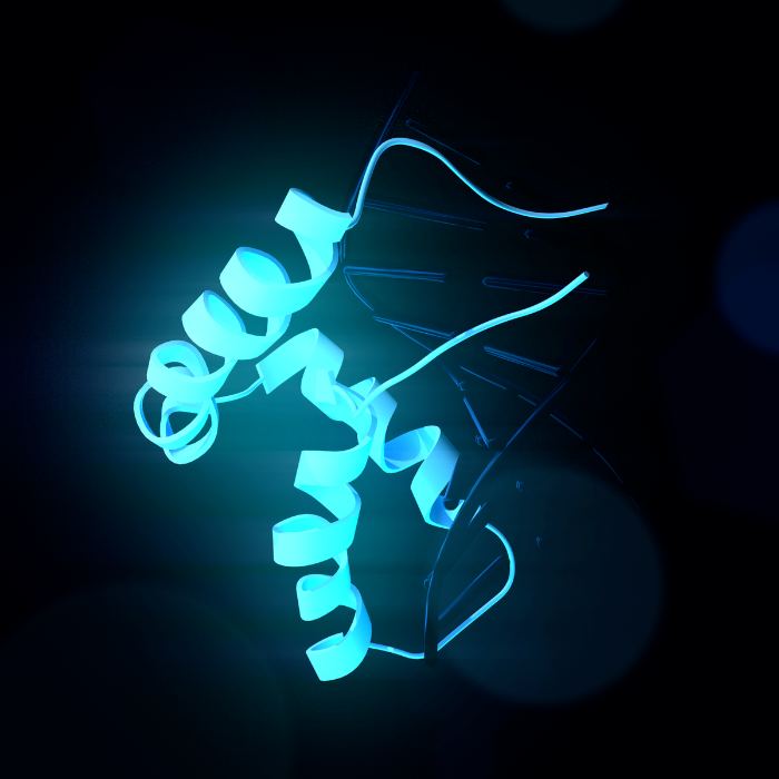
Two viral proteins, E6 and E7, are produced by the infected cells. When bound to a particular cell protein, E6 triggers that protein to label the tumor suppressor gene P53 for destruction. Consequently, the cell is deficient in a key component for preventing mutations and cancerous transformations. Another viral protein, E7, is responsible for cell death by “hijacking” a cellular protein engaged in crucial regulatory mechanisms. This causes the release of transcription factors that actively promote cell multiplication. Two proteins assist the virus in disrupting the cell’s basic metabolic and developmental processes, leaving the cell vulnerable to mutations.
However, this is by no means the sole mechanism by which HPV causes cervical cancer. In fact, new mechanisms are disclosed virtually every week. These two, though, are the sharpest and most consequential.
The dirty dozen
Researchers have had a hard time finding new tumor-inducing viruses due to the intricacy and variety of the processes and regulatory systems already engaged. This is one of the reasons why just a handful of anti-cancer drugs that target viruses have been shown to be effective in people.
Epstein-Barr virus (EBV) for nasopharyngeal cancer and Hodgkin’s lymphoma; hepatitis viruses B and C for liver cancer; human T-cell lymphotropic virus type 1 (HTLV-1) for cervical cancer; and human herpesvirus 8 (HHV-8) as a trigger for Kaposi’s sarcoma are all well-established viruses that cause cancer in humans. Polyomavirus and hepatitis C for non-Hodgkin’s lymphoma, and an unidentified agent for a kind of pediatric leukemia, remain on the list of possible tumor-causing viruses. The tally of cancer viruses in humans that have been “condemned” continues to increase today.
Helicobacter pylori and stomach cancer
Several kinds of cancer in humans now have a known link to infections with certain viruses, bacteria, and parasites. 15–20% of all malignancies are believed to have an infectious cause. But we weren’t always aware of the cancer-causing bacteria. In fact, this medical dogma was toppled anew by a dogged scientist, a little bit of self-experimentation, and a giant leap forward. The Australian scientist Barry Marshall, his colleague Robin Warren, and the rod-shaped bacteria Helicobacter pylori are the story’s primary characters.
In 1982, the medical community accepted the dictum that “Bacteria are everywhere—but the human stomach is bacteria-free.” Because of the very acidic conditions present in the stomach, it was often believed that no bacteria could thrive there. And that was not all. The majority of medical professionals and scientists in the field of pharmacology, as well as patients, attributed the pain of gastric mucosal inflammation and subsequent gastric ulcers to the presence of acid in the stomach. The pharmaceutical industry promoted acid blockers of different sorts with the slogan “No acid, no ulcer,” and sales were booming as a result.
Its home is the stomach
Pathologist Robin Warren found something that shouldn’t be there when he examined tissue samples from stomach ulcers and irritated stomach mucosa under a microscope: bacteria that are full of mucus. And that was the case in over half of the samples he tested. The number of these cells seemed to be correlated with the degree of inflammation. Warren consulted with doctor Barry Marshall after concluding that his finding must be significant.
As a team, they achieved the first successful cultivation of the bacteria. They could now undertake microbiological experiments to learn how the bacteria manage to thrive despite the harsh circumstances of the stomach. To begin attaching to the mucosal cells, bacteria first use their flagella to dig deeply through the protective mucus layer. Meanwhile, they used an enzyme called urease to convert the uric acid produced by the cells into carbon dioxide and ammonia. This created a neutral environment that bypassed the stomach’s defense against microbes.
A sip alters the course of history
However, these results did not establish beyond a reasonable doubt that this particular bacteria caused an inflammation or cancerous tumor. Because the conclusive evidence was still absent. In July of 1984, after months of testing, Marshall decided to take a daring step: he began testing on himself. On the informal principle of “a small sip for a person-a big sip for medicine,” he consumed a brackish solution containing billions of germs. As Marshall reflected, “Everyone said, you’re crazy. It seemed like too incredible an explanation for something as complex as stomach ulcers.”
In a matter of days, he started feeling sick, like he had the flu, and in two weeks, he had gastritis. With the use of a tissue sample, he was able to see that his mucosal cells were covered in a thick layer of germs. Thus, Marshall successfully exposed Helicobacter as the true culprit in the inflammatory process. At that time, though, the link he suggested between the bacteria and stomach cancer had not been proven.
The murderer’s in the blood
However, since then, his theory has been independently verified: antibodies may indeed identify the bacteria in the blood. Three further significant epidemiological investigations followed this. Scientists looked for Helicobacter antibodies in the blood of thousands of stomach cancer patients, whose blood was collected and preserved decades ago as part of earlier research, and compared the findings to the values of a similarly large sample of patients who did not have cancer of the stomach.
Those who were infected with Helicobacter 20 years ago had a six-fold increased risk of developing cancer compared to those who were now free of Helicobacter. These findings were so compelling that in 1994, the World Health Organization’s International Agency for Research on Cancer (IARC) officially labeled Helicobacter pylori a class 1 carcinogen, the first identified bacteria capable of causing cancer.
Terms related to viruses, cancer, and genes
Most of what we know about how genes play a role in cancer was uncovered via studies of tumor viruses. The relative simplicity of the viral genome has been the only thing that has allowed researchers to identify, or at least narrow down the DNA building blocks involved in the formation of human cancer.
The link between viruses and cancer has been recognized to exist in animals since the middle of the past century, but only in people since the 1980s. Unlike animal tumor viruses, however, the link between human cancer viruses and the disease is based on correlations or presumed processes rather than a clear cause-and-effect premise. The viruses have a role as either cofactors or cocarcinogens in this scenario. This demonstrates undeniably that the process of carcinogenesis in humans is intricate and multi-staged.
At least six families of DNA viruses and two families of RNA viruses are suspected in the development of cancerous human tumors. Among the examples are Retroviridae and Flaviviridae as RNA virus families and Hepadnaviridae, Herpesviridae, and Papillomaviridae as DNA virus families.
RNA viruses:
Those are the viruses that employ RNA rather than DNA to store their genetic information. Single and double-stranded forms of the RNA are possible. Examples of RNA viruses include influenza, polio, and Ebola; the hepatitis C virus has been linked to cancer.
Retroviruses:
The retrovirus family is also an RNA virus. To integrate their genome into the host cell’s chromosome, retroviruses employ the enzyme reverse transcriptase to create a DNA translation of their genome. Here, it is incorporated into the DNA of the cell for good. The AIDS virus and HTLV-1, a virus linked to leukemia, are examples of such pathogens.
DNA viruses:
These viruses have their genetic material in the form of DNA and may thus integrate it into the host cell’s genome without first requiring “translation.” Herpes viruses, human papillomavirus (HPV), linked to cervical cancer, and Epstein-Barr virus, linked to certain types of leukemia, are all examples.
Transduction/transformation:
This is the process through which healthy cells mutate into cancerous ones. This mechanism has been greatly elucidated by the capacity of viruses to convert cells in culture. The altered cells display hallmarks of cancerous tumors, including unchecked multiplication, reduced reliance on supporting tissue, and less need for external growth hormones. When transplanted into a new host organism, some altered cells may potentially become cancerous.
Tumor virus transformation of a cell always exhibits the following three characteristics: it is a “one-hit” process, meaning that the transformation is triggered by a single viral infection of the cell. In addition, the cell retains some viral genetic information, and the transformation is maintained by the continued production of a subset of viral genes.
Oncogene:
A gene that has a significant role in causing or facilitating the progression of cancer. Point mutations, rearrangement of its DNA building blocks, loss of individual segments, and excessive, uncontrolled gene expression triggered by viruses all contribute to its activation and transformation into a tumor gene. The Greek term for “mass” or “lump” is whence we get our modern word “onco.”
Cellular Oncogenes:
An oncogene may mean either a normal gene that has been changed to become active in tumor cells or a cellular gene that has been activated in tumor cells.
Viral Oncogenes:
DNA viruses may cause cancer because they include a gene that isn’t found in cells but has the potential to do so when infected, or because they contain a version of a normal cellular gene that has been changed or activated to operate as a tumor inducer.
Proto-oncogene:
A precursor of both viral and cellular oncogenes, a normal gene of the cell that may be turned into an active oncogene by external factors.
Tumor suppressor gene (TSG):
Genes encoded by a cell’s own DNA inhibit cancerous transformation. These genes may have a bigger effect on carcinogenesis than the active oncogenes.
Woolhouse M, Scott F, Hudson Z, Howey R, Chase-Topping M (2012). “Human viruses: discovery and emergence”. Philosophical Transactions of the Royal Society of London. Series B, Biological Sciences.
They all want blood, whether it’s Dracula, Lestat, or Nick Knight. An eternal mystery, the legend of blood has long captivated the human race. Numerous civilizations have unique myths and stories about the crimson elixir of immortality. “Blood is a very unique fluid,” Mephistopheles said. Therefore, he insisted Faust use a “drop of blood” to seal the pact. But what makes blood so special?
Science, following in the footsteps of religion and literature, has endeavored to unravel the mystery of the life force that courses through our veins.
Death is an inevitability when blood loss is excessive. The question then becomes why blood is so important and what role it plays in the human body. Our blood volume is between 5 and 6 liters. So where does the blood flow originate from?
Transferring oxygen from the lungs to the body’s numerous tissues is the blood’s primary role. The carbon dioxide is transported back to the lungs on its return journey. Blood does play a role in gas transportation, but it is not the only mechanism. It serves as a highway for many other compounds to reach their destinations in the body. For instance, hormones fall under this category, but so do essential nutrients for metabolic function.
Toxic waste products are carried away from the body by the blood to the liver and kidneys, where they are eventually eliminated. And it’s vital to the body’s ability to fight against invaders.
Blood of myth
As it relates to rituals, religions, and supernatural
There is a common thread connecting myths and religions across the globe, and that thread is the story of blood. Homer likely has the first account of a blood-related magical ritual. On his way to meeting the spirits of the dead, Odysseus must first sacrifice a number of animals. The only way the deceased can remember the hero’s name and face again is if they consume this elixir.
Vampire mythology certainly isn’t exclusive to the West. The Germanic peoples, the Romans, the Celts, and even the Africans all had ideas about the undead slurping off human blood.
They were called “Lamias” by the ancient Greeks. Women who died in mysterious circumstances were said to be resurrected as vampire-like ghosts. They devoured the victims and drank their blood.
However, blood has always played an important symbolic role in both literature and religion. Both the ancient Chinese and the Egyptians had the belief that consuming the blood of a murdered enemy would bestow upon the drinker the same supernatural abilities as the deceased. Legend has it that Germanic peoples used the still-warm blood of slaughtered bears, wolves, and cattle as a currency to purchase these animals’ territories.
Killing for the sake of the blood
Some religions practiced blood sacrifices in which human or animal lives were sacrificed as an act of worship. Blood was worshiped as a deity’s beverage among the Mayans. Because of this, it was necessary for there to be a constant flow so that communication with the gods and ancestors would be maintained.
Ancient Egyptians and Babylonians shared this view, thinking of blood as the conduit between the body and the afterlife. Even in the Old Testament, blood is connected with life. During the Inquisition, lamb’s blood was painted on walls to ward off bad spirits and as a reminder of the atoning power of animal sacrifices. However, at the Lord’s Supper, we partake in the symbolic drinking of Christ’s blood for the purpose of receiving forgiveness for our sins.
For a long time, blood was also a common way to symbolize intimate relationships. For instance, the term “blood brotherhood” refers to the unique closeness shared by two buddies that normally exists only between biological brothers.
Blood, along with phlegm, black bile, and yellow bile, was thought to be one of the four lifebloods back in the Middle Ages. They believed that disturbances in the equilibrium of these four humors were at the root of all diseases. Excess blood has to be drained to restore equilibrium. However, until the nineteenth century, physicians still routinely performed bloodletting as one of the most prevalent therapies for any and all ailments, even after the four-fluid hypothesis had become obsolete.
The discovery of blood circulation
William Harvey.
For a long time, little was understood about blood, despite its central role in literature and religion. What exactly is blood composed of? What causes its structure to take shape? These mysteries were solved years later by diligent scientists. Up until that time, the common belief was that food was used to make blood, in accordance with Galen’s teachings.
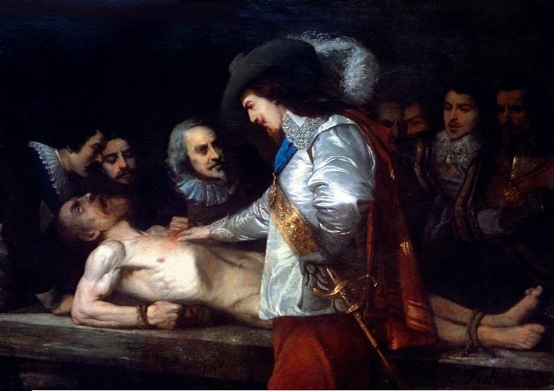
British physician William Harvey made the discovery of blood flow in the first part of the 17th century. The blood needs a propelling device, such as a pump, to prevent it from pooling in the legs due to gravity. The heart is responsible for carrying out this process. Blood can only flow one way through the heart, and the heart valves make sure it’s out of the body and away from the heart in the arteries and back in via the veins.
But how can we explain the constant circulation of blood? Instead, the blood flow should slow to a near stop between contractions before being pushed forward once again. Blood arteries are no longer hard tubes, thanks to nature’s solution of making them stretchy. So, the veins close to the heart get more blood between heartbeats and then release it all during expansion. The blood is essentially discharged in a wave form from the heart before being transformed into a steady flow.
The individual cells found in the blood
Blood is made up of separate cells. If you scratch your finger and look at the drop of blood under a microscope, you will discover that blood is not a homogenous liquid. A few are white, while the rest are bright red. Because blood is both a fluid and an organ, every component has a unique purpose. It’s not only a conduit for oxygen and nutrition, but also for waste products like carbon dioxide and chemicals.
This is where your body’s first line of defense stands against invading cells and organisms. The “juice of life,” then, contains a cast of characters. White blood cells, such as B cells, T cells, and natural killer cells, act as “soldiers” and “guard troops,” mingling with the “pure transport workers” like hemoglobin, which carries oxygen.
Red blood cells (erythrocytes)
Erythrocytes are the primary component. The form of a red blood cell is similar to an egg’s, albeit it is somewhat flatter in the center. Hemoglobin, an iron-containing protein that gives blood its red hue, is responsible for carrying oxygen to the body’s tissues. During this step, the heme groups’ iron molecules bind with the oxygen molecules.
Reduced hemoglobin levels lead to the condition known as anemia. There are a number of possible reasons for this anemia, but in the Western world, a deficiency of iron in the blood is the most common. Iron deficiency is more common in women because of menstruation, although iron-poor diets (such as those followed by vegetarians) may also lead to anemia.
White blood cells (leukocytes)
The blood contains both red and white blood cells. Disease prevention relies on the immune system, which includes white blood cells. They are not only present in the blood, but also in the lymph. Antibodies are produced by B cells, which subsequently bind to any foreign substances, such as viruses or bacteria, that have entered the body.
Upon detection, so-called T-helper cells signal for the elimination of these binding complexes. When a virus is the offending agent, T-helper cells dispatch killer cells to do away with the contaminated tissue.
Platelets (thrombocytes)
Despite popular belief, there are more components to blood than just red and white blood cells. If a vein or artery were slashed, blood would spurt out in all directions, and the victim would bleed to death. Platelets’ function is to stop this from happening. They start blood coagulation by closing up damaged blood arteries.
Maintaining a consistent concentration in the blood is essential for their functions. Bleeding may occur if there are not enough thrombocytes in the blood, and thrombosis can develop if there are too many. It’s the formation of clots in the blood vessels that prevents the normal circulation of blood.
Blood plasma
Blood cells and thrombocytes still need a liquid medium to move across the body. Plasma in the blood does this work. It consists of almost entirely water and has a 96% water content. Immunoglobulins, which transform into antibodies after an immunological response, are part of the four percent of plasma proteins.
Blood coagulation
Healing of bare cuts by themselves
Humans have the remarkable ability to swiftly create a scab over any cut or scratch. This scab is made entirely of dried, coagulated blood.
It stops bleeding from broken blood vessels. In the normal course of events, platelets produce a hemostatic plug a few seconds after damage, which is then strengthened by a tiny network of fibrin fibers. Fibrin acts as a hemostatic and initiates blood vessel healing. The blood does not naturally contain it, but rather it is generated via a series of precursors involving over 30 distinct elements before it can be found there.
Liver dysfunctions are particularly dangerous for blood clotting since the liver is responsible for producing several of the clotting components. In reaction to damage, clotting is initiated by signals sent only locally. The ultimate goal is to keep the blood from clotting entirely, since this would render it useless as a transport medium.
However, there is no guarantee that bleeding will cease after an injury. If you look at leeches, for instance, they can draw five times their body weight in blood in half an hour without the bite wound clotting. Their method is to secrete hirudin into the wound while they feed, which keeps the blood supply so the leech can keep sucking. Hirudin binds to and neutralizes thrombin in the circulation, preventing it from initiating a clot.
Patients are already being given hirudin and related medicines to prevent thrombosis after surgery or during long-distance flights.
Nonetheless, chemical compounds aren’t the only things that may stop blood from clotting. Some individuals are born without this defense system functioning properly. They have hemophilia, a disorder that runs in their family. When an injury develops, no scab will form, and the wound will continue to bleed unchecked, perhaps causing a lot of blood loss. Most notably, members of European royal families have been affected by the inherited bleeding disorder hemophilia.
Hemophilia
In late 19th-century England, Queen Victoria married her daughters into the most powerful governing houses on the continent in an effort to solidify her position there. Soon, babies were born. However, many of them, particularly male kids, had to overcome a severe disability: Whenever they were hurt, their blood poured non-stop. On top of that, the wounds just weren’t healing. Among them, only a few grandchildren of Queen Victoria reached adulthood. So many of them died either soon after birth or even before they turned 18.
So how did this mysterious illness that mostly targeted males rapidly spread among Europe’s royalty (kings, dukes, etc.) over such a huge geographical area? The sick, power-mad queen was actually the source and the original cause of this disease.
The mutation of an X-chromosomal gene encoding a blood clotting factor is identified as the root cause of so-called hemophilia. It is generally accepted that males have just one X chromosome and females have two X chromosomes. Due to its recessive inheritance pattern, the condition does not manifest itself if the second X chromosome’s intact gene can generate a functional clotting factor.
Because of this, women who have the abnormality on just one X chromosome are not afflicted with the illness but are carriers. However, there are no clotting factor genes on the Y chromosome in men. Therefore, if the X chromosome gene is faulty, it cannot be substituted by a normal gene on another chromosome.
The son of a healthy mother who is a carrier has a 50% chance of inheriting the faulty chromosome and the sickness from her. This is because males obtain their X chromosomes from their mothers. To develop the condition, a female must inherit a faulty chromosome from both her parents. If this is the case, however, it means that the father is a hemophiliac as well.
Hemophilia was uncommon in women since male hemophiliacs seldom achieved sexual maturity, particularly in the past. The number of women who are infected with HIV has grown, despite the availability of improved treatment alternatives. Even though a cure has not been found, the illness may be managed with frequent injections of the clotting factors.
The blood group system
Medical professionals always insist on finding out a patient’s blood type before administering a transfusion, no matter how dire the situation is. Just how crucial is it that the patient obtain blood from someone with the same blood type as them? Because of the high mortality rate prior to the identification of blood groups, blood transfusions were only sometimes carried out. Due to this, they were outlawed in several European nations around the turn of the last century.
However, Viennese physician Karl Landsteiner (1868–1943) wondered if there was a pattern to the transfusions’ inconsistent performance. He combined his own blood with the blood of five coworkers in a wide variety of ways. His work on this led to the development of the blood group system in 1901, and he was eventually given the Nobel Prize for his efforts.
Blood groups A, B, and 0 serum include “clumping proteins,” which bind to red blood cells of other blood types and cement them together, causing a blockage in the blood arteries. The antigens on the surface of red blood cells are the proteins that are there. Erythrocytes that have a distinct surface make up are identified as invaders and attacked. The key-lock mechanism makes this possible. Specific antibodies are produced by lymphocytes, which then bind to foreign antigens to create an insoluble complex.
Rhesus factor
Landsteiner’s research greatly assisted in the care of injured servicemen during World War I, among other areas of medicine. In spite of this, he sometimes encountered obstacles beyond explanation, compelling him to delve further into the crimson elixir of life’s mysteries. It wasn’t until 1940, when working with rhesus monkeys, that he realized there was still another factor in compatibility. This rhesus factor was discovered by isolating a previously unknown component of the red blood cell membrane.
Having or not having (Rh+) is the underlying premise (Rh-). To what end, however, does the rhesus factor serve as a proxy for blood safety? Hemolysis may occur in a patient with an Rh-negative blood type if a donor with a positive Rh factor is used. This results in the complete elimination of RBCs (red blood cells) from the blood.
When a patient receives donor cells, their own immune system rejects them because the patient’s cells lack the rhesus factor. That is, they generate anti-rhesus factor antibodies. A group of immune cells gets a message from the antigen-antibody complex that an invading pathogen must be dealt with. Specialized T-killer cells eliminate the complex by destroying it. Hemoglobin is removed when the cell membrane has been destroyed. The opposite is not true, though. Therefore, Rh+ individuals are safe to receive blood transfusions from Rh- donors.
Potential dangers to the unborn
Furthermore, the Rh factor is crucial during pregnancy. Generally, things go well with the firstborn. A woman who is Rh- but has given birth to a kid who is Rh+ will have antibodies in her blood that attack the Rh+ cells. If she conceives another kid who is Rh+, the antibodies will cross the placenta and cause hemolysis in the baby.
Rh incompatibility used to be a leading cause of stillbirths. To prevent the immune system from developing defense cells against the child’s blood, doctors now vaccinate mothers with anti-Rh antibodies right after the birth of the first child. This vaccination is given again in the second and third trimesters of pregnancy.
Twenty distinct blood types have been discovered in humans. The AB0 and Rhesus systems are two of the most popular.
Risks of blood transfusion
The curse of blood
Unfortunately, blood carries more than just hormones, oxygen, and nutrients. Once within the body, pathogens may travel through the bloodstream to other organs with equal efficiency.
Although blood transfusions save lives, they pose a danger if the blood provided is not adequately screened for infectious agents and compatibility. This has been recognized at the very least since the HIV-contaminated blood crisis of the 1980s. Standard testing for HIV and hepatitis viruses, for instance, did not become accessible until the late 1990s.
Hepatitis
In the late 1970s, after giving birth, around 7,000 women received an anti-D immunoglobulin vaccination in one country to protect against rhesus factor incompatibility in future pregnancies. However, roughly 2,500 women got hepatitis C because the virus was present in tainted batches.
Meanwhile, hepatitis infection during transfusions is less likely due to systematic testing of blood supplies. Infections through tainted blood units happened often before dependable diagnostics for hepatitis viruses were available. Because it might take ten to twenty years for normal symptoms like fever and jaundice to manifest, many blood donors are unaware that they are carriers of hepatitis viruses.
Then, if the illness does spread, there are many important stages it might take. In most people with hepatitis B, the illness clears up on its own when the body develops antibodies to combat the virus. However, chronic development occurs in 5 to 11% of instances. Patients also generate some anti-hepatitis B antibodies, but not nearly enough to drive the virus out of liver cells.
Hepatitis C poses a much greater risk since, unlike hepatitis B, there is currently no vaccination available to prevent the disease. Liver cirrhosis occurs in severe instances. After successfully cultivating the hepatitis C virus in cell culture, scientists still have high hopes of discovering new treatment options.
West Nile Virus
According to legend, ravens began dropping dead from the skies just before Alexander the Great’s death, a sure sign that he had contracted the West Nile virus. In 1999, a similar event happened in New York City’s Central Park. The virus had flown over the Atlantic to the West, where it quickly disseminated throughout the Americas. The virus has also been a recurrent topic of discussion in the European media.
The West Nile Virus was first discovered in 1937 in Uganda’s West Nile District, hence the name. Birds are the primary hosts, and they may be found in both tropical and temperate regions. However, mammalian species, including humans, are rapidly becoming its victims. More than 4,000 cases were recorded in the United States in 2002, with 284 deaths. Blood transfusions were the source of infection in 13 instances.
The Culex pipiens mosquito is the primary transmitter of the West Nile virus. Scientists pondered the mystery of the virus’s rapid spread across the Atlantic. More research into the mosquito species led scientists to discover that two subspecies call Europe home. One likes to bite people, while the other likes to bite birds. However, an intermediate variety that bites both birds and people in the Americas makes for an efficient viral carrier.
At first, it was thought that the virus could not spread from person to person. But since the beginning of the millennium, evidence has accumulated to support the idea that it is likely spread by the blood during transfusions and organ transplants.
Avoiding unnecessary risks requires a few weeks of the waiting period after returning from North America between June and November before a donor may give blood again. Epidemics tend to happen in the summer because the disease and its carriers both multiply more quickly when temperatures are higher.
West Nile encephalitis brain tissue courtesy of the Centers for Disease Control and Prevention
The majority of infected people show no symptoms, but those in the 20 percent who do have flu-like symptoms. Danger arises when the virus causes meningitis by crossing the blood-brain barrier. No effective cures still exist at this time.
Fighting malaria with sickle cell anemia
Life-saving genes that are also deadly
There are moments when nature seems to follow its own peculiar set of rules. That’s what the researchers who identified the relationship between malaria and sickle cell anemia must have believed. The latter is an inherited disorder brought on by a change in the hemoglobin gene. This causes the red blood cells to take on an abnormal sickle form, which causes them to get lodged in the arteries and prevent oxygen from reaching the affected organs. A heart attack happens when blood arteries get clogged and no longer provide blood to the heart.
Darwin’s “Survival of the Fittest” theory predicts that harmful mutations should be rare and eventually eliminated by natural selection. The prevalence of sickle cell anemia in malaria-endemic areas like West Africa is a curious situation. Scientists discovered that the altered cells provide a selective advantage in some environments. The malaria parasites only target RBCs. However, sickle cells are more susceptible to destruction, leading to the simultaneous death of both the infections and the host cells.
Deadly mosquitoes spread disease
The mosquito-borne illness malaria is well-known across the world. The Anopheles mosquito is responsible for spreading the parasitic protozoan Plasmodium. When a mosquito bites an infected person, it takes in the plasmodia along with the blood. Transmission from one victim to the next takes place by physical touch.
Once inside a host, parasites will go toward the liver to multiply. Once within the red blood cells, the daughter cells grow rapidly before being ejected in a violent burst. As a consequence, malaria is associated with recurrent fevers, for which it is recognized.
There are two phases to the malaria parasite’s life cycle: the mosquito-borne sexual cycle and the human-borne asexual cycle. Female and male plasmodia fuse in the Anopheles mosquito. The so-called sporozoites form in the egg between eight and sixteen days after fertilization and then enter the bloodstream of a human host during the subsequent sucking cycle.
They develop into schizonts in the liver cells, the subsequent intermediate stage. Plasmodium falciparum (Malaria tropica) is the most severe form of the disease, in which all schizonts grow into cells whose daughter cells damage liver cells and then infect erythrocytes in the blood.
Only a subset of schizonts reaches this stage of development in the mild and harmless malaria forms. The remainder goes into a hibernation-like state in the liver, where it may remain viable for a long time. In time, they develop into adults and cause the condition to recur.
The parasite is very well-protected since it is able to evade immune system assaults by hiding out within the host’s liver and blood cells at all times. Furthermore, it generates a protein whose composition intermittently changes, making immune system adaptation difficult.
Expectations for a vaccine
The Plasmodium and Anopheles genomes were first sequenced by a group of scientists in 2002. Antimalarial medications exist, however, malaria parasites rapidly evolve resistance to these treatments.
As of 2022, the RTS,S, also known under the trade name Mosquirix, is the only malaria vaccine that has been licensed for use anywhere but the European Union. Developed in 1987, RTS,S received WHO approval for “wide usage” in children only in 2021.
Attacks on the immune system
The immune system is to contemporary people what the heel was to Achilles. It keeps harmful bacteria at bay, but when it breaks down, disaster strikes. Specifically, the white blood cells are the “wonder spot” because they are the first line of defense against infections. The consequences of their destruction or damage are catastrophic. Disabling the immune system leaves the body vulnerable to pathogens it would normally be able to fight off.
AIDS
This is the root of the AIDS epidemic, which has been likened to the Black Death in Europe in the 14th century. They are working around the clock to stop the spread of the illness, which is affecting Africa particularly badly.
The HIV virus is responsible for the disease; it is a kind of retrovirus whose genetic material is encoded in RNA but becomes fixed as DNA inside the host cell’s genome. When a virus integrates its genetic information into a host’s own, it becomes very tough to eradicate. It can only reproduce in the presence of CD4-positive cells throughout the body.
It is the T-helper cells that are principally responsible for cellular immunological defense in humans. Following the integration of viral DNA into the helper cell’s nucleus, the cell is coerced into accepting new instructions. The T helper cell is no longer producing immune response-coordinating messenger molecules, but rather HIV viruses, which eventually cause the cell to die. As a result, the released HIV viruses infect and hijack more T helper cells.
During this period, the body’s immune system is constantly fighting off infections. About ten billion new viruses are created each day by infected T-helper cells, and one billion T-helper cells each day die and need to be replaced. At first, this works well, but eventually, your body just can’t make enough T-helper cells to replace the ones you lose every day. The patient is now open to infection from any germ since their immune system is no longer functioning.
After around 10 years, the decreased immune system might cause a variety of illnesses. It is generally accepted that AIDS sets in when the CD4 cell count drops below 200 per microliter. What this implies is that the body is open to contracting deadly infections. Thanks to modern medical knowledge, we may put off this process and save ourselves the trouble it might bring. Unfortunately, there is currently no hope for a cure.
Hope for a cure
Vaccine development for HIV is still the primary focus of ongoing studies. Protein molecule Gp120 is the entry point for HIV viruses into infected immune cells. Gp120 changes its structure in response to binding the CD4 receptor. Knowing how gp120 changes shape leads scientists to suppress HIV by employing medicines that impede the shape shift. But as of 2022, HIV is still not curable but only controllable with HIV treatment.
HIV may be transmitted by any kind of skin-to-skin contact that results in a blood transfusion. Blood supply contamination has been a major concern for quite some time. However, in the meantime, testing blood donors for HIV has become routine. It takes many weeks after infection for antibodies to build in the blood, so there is still a certain residual danger. After that, the HIV test is able to detect them.
White blood cells
When compared to the decline of lymphocytes seen in AIDS, the other extreme is seen in leukemia, when an unchecked proliferation of one or more kinds of white blood cells occurs in the bone marrow, but these cells are non-functional. The progenitors of mature lymphocytes are targeted by acute lymphocytic leukemia, whereas granulocytes are the primary target of myeloid leukemia. DNA mutations in the stem cells are the cause of the deterioration.
The mutated cells circulate around the body and eventually end up in different organs. In addition, the cluster of leukemia cells in the bone marrow pushes out the stem cells that normally give rise to all the other types of blood cells. This causes abnormal hematopoiesis, which leads to a shortage of blood cells over time. Weakness, pallor, an increased vulnerability to infection, and a propensity to bleed are all common signs of leukemia.
Current treatments for leukemia include chemotherapy and stem cell transplants. Chemotherapy is the use of strong medications to halt the development and division of cancer cells. These cytostatic medications are effective against cancerous cells, but they can harm healthy organs. Subsequently, patients often have awful adverse effects such as vomiting, mucosal irritation, and alopecia.
A frantic hunt for potential treatments
Because of the inherent dangers of bone marrow or stem cell transplantation, new therapeutic approaches are desperately required. Scientific efforts are presently concentrated on pinpointing optimal targets.
The enzyme DOT1L is an example of a gene activator that plays a crucial role in leukemia development and hence is a possible target for novel medicines. DOT1L limits the activity of the DOT1L enzyme, paving the way for killing the afflicted cells of leukemia patients.
The killer cells of the immune system are the topic of study for experts. Scientists are able to grow in the lab killer cells capable of killing leukemia cells thanks to genetic changes. Motherwort has a compound that kills leukemia cells as well. In this method, normal cells are protected, unlike with present pharmaceutical options. Leukemia is often treated with chemotherapy. The leukemia cells are chemically destroyed by the drugs.
The cause of leukemia remains a mystery after years of study. According to the most widely accepted theory, leukemia develops when certain types of blood cells acquire mutations in their DNA.
Those who have been exposed to ionizing radiation, whether from the sun, X-rays, or radioactive sources, are at a higher risk. However, cytostatics, benzene, other chemicals, and immune system abnormalities may all play a role in bringing on the condition. A hereditary component cannot be ruled out.
Artificial blood
The safe alternative
Donating blood can save lives. Summer is particularly prone to bottlenecks caused by a lack of blood supplies. More blood is required, on the one hand, since accidents are more common among travelers. On the other hand, donors tend to stop donating while they’re away.
However, there are several factors at play, such as the aging of the population, that contribute to the overall decrease in blood donors. Surgical treatments, such as hip replacement, are becoming more common among the elderly as a result of the expanding capabilities of modern medicine. More blood must be stored for this reason.
However, the average lifespan of a blood unit is about 40–42 days. Donated blood is becoming less effective because of the rising prevalence of blood-borne viruses like HIV and hepatitis. Many individuals have been wary of giving or receiving human blood since it was discovered that blood transfusions might spread illnesses like AIDS and hepatitis.
Seeking a substitute that fits
Because of this, doctors are looking for alternatives all the time. The next best thing would be synthetic blood. After decades of research, however, experts conceded that it would be too hard to create a fluid with all the characteristics of human blood. Therefore, scientists are focusing more on two crucial activities, oxygen transportation, and blood loss compensation.
Scientists have come to believe that synthetic blood should be thicker than real blood in order to limit the danger of capillary breakdown. In order to counter this, scientists in the United States have created a format called MP4. Specifically, this makes use of RBCs (red blood cells) extracted from human donors’ blood that have been frozen.
Polyethylene glycol is added to the blood to make the hemoglobin molecules bigger, making the blood thicker. Moreover, MP4 has a stronger affinity for oxygen than other products, so the oxygen is released only in the capillaries, where it is absorbed directly by the surrounding tissue. In theory, artificial blood doesn’t care about your blood type.
On paper, artificial blood is more efficient in its primary function—oxygen transportation—than “real” blood. However, since artificial blood loses 50% of its oxygen-transport ability after a few days, it can currently only be employed for acute applications. But in contrast to donated blood, artificial blood can be kept for a year or longer. Natural blood, on the other hand, has a shelf life of just one month before it deteriorates.
How about hemoglobin extracted from animals?
To anybody who has ever been on the mudflats, the little mounds of sand left behind by the lugworm are instantly recognizable. But who knew that these unassuming creatures would provide a fresh ray of hope in the fight against disease? The worms’ hemoglobin might one day replace human hemoglobin or be used to preserve organs.
The protein hemoglobin in human blood can store four molecules of oxygen. Lungworms’ hemoglobin protein holds 156 oxygen molecules. The lugworm is also a universal donor since its blood lacks the A, B, and O antigens that distinguish different types of human blood.
Pigs’ red blood cells also have been used to successfully create artificial blood. This type of artificial blood has very large molecules for carrying oxygen with characteristics that are similar to those of human blood.
Moreover, when scientists need to boost the blood volume due to blood loss, they add in plasma expanders. This is an ideal substitute for blood. As scientists believe, nothing currently stands in the way of the industrial manufacture of artificial blood.
The inability to sleep leaves you feeling drained the next day. If there weren’t any particularly stressful events the day before, it might just be the mattress giving you trouble sleeping. This is the case even if it retains the ideal combination of hardness and flexibility. Because the metallic components of mattresses, particularly innerspring mattresses, are thought to have a detrimental effect on people. A magnetization of the mattresses or an amplification of electrosmog by the utilized metal could be to blame for this. But, is this just a superstition? Do innerspring beds and their “high” EMFs (electric and magnetic fields) have a negative health impact on people?
Metal components are subject to electromagnetic field changes in their surrounding environment, including both increased and reduced field strengths. Coil springs, however, do not have much of an impact on the levels of electromagnetic fields in the bed. As soon as you are lying down in bed, the innerspring metal’s relatively weaker electromagnetic effects are immediately overridden by the much bigger human body.
Mattresses’ potential to boost the strength of electrical radiation is minimal at best.
The Earth’s magnetic field is altered by innerspring mattresses
Then, might it be that the metal in innerspring mattresses interferes with Earth’s magnetic field? The innerspring of a bed and any nearby sheet steel or screws distort the magnetic field around the bed. It’s easy to demonstrate this effect by using a compass. Cardinal directions are elusive in many structures due to the presence of steel parts. The innerspring mattress contributes negligibly to these shifts in this setting.
But do innerspring mattresses pose any health risks or interrupt sleep quality? The present level of scientific knowledge suggests that weak magnetic fields pose no threat to our health or the quality of our sleep. This is equally true with respect to electrical radiation.
The sleep laboratory studies on people exposed to radiation find either no alterations or, in fact, a really good influence on sleep. Both middle-aged men and younger men, as well as middle-aged women, experienced this effect. Electrosensitivity as a concept also lacks demonstrable evidence.
Optimal sleep quality is determined by your physical environment
People consequently give far greater weight to a different factor when it comes to sleep quality: the wide variation in the mechanical, thermal, and diffusion qualities of mattresses. These physical factors are crucial for a peaceful and rejuvenating night’s sleep. The ideal mattress combination is based on personal circumstances and preferences. And this is all the same whether with or without an innerspring core.
From a sleep medicine perspective, an adjustable mattress—the one that adapts to your lying position—is more significant for health than the little risk of electrosmog possessed by innerspring mattresses. One piece of advice the researchers have is to make sure the bedroom is never too hot or too humid for the person sleeping there.
I had my first child, and now my shoes don’t fit anymore! Many women appear to have this problem: During pregnancy, their feet seem to have grown. But is that scientifically true or are women just imagining it? This question once became the subject of a doctoral thesis. Physician Johannes Christian Hentschel regularly measured the feet of 40 expectant mothers during pregnancy. The research showed that pregnant women’s feet did get bigger in length, width, and size.
On average, their foot became 1.8 mm longer and 2 mm wider during pregnancy. This increase in length corresponds to about a quarter of a shoe size, the doctoral thesis says. And the right foot grew more than the left.
So it’s obviously not imagination. Towards the end of pregnancy, the phenomenon occurred to a greater or lesser extent in almost all women. 70 percent of them still had shoes that fit, 30 percent needed a larger shoe size or had to put on shoes that were cut a little wider.
More water in the feet
The feet of a pregnant woman get larger because the body retains extra water in the tissues. As the uterus grows, it puts pressure on lymph vessels and veins in the pelvis. Because of the increased pressure in the veins, blood from the foot flows more slowly back to the heart, causing backlogging. This causes more water to seep from the blood and get trapped in the tissues. This is most visible in the feet due to gravity.
Pregnancy hormones further facilitate this growth in the foot size by relaxing the tissue to allow the uterus to expand. However, this also facilitates water transport from the circulatory system into the tissues Compression stockings are recommended for pregnant women who have this fluid retention. They allow less water to be absorbed by the tissue due to the external pressure put on the legs. Although the compression stockings do not stop the feet from growing during pregnancy, they slow the process down significantly.
Usually just a passing phase
Thankfully, these alterations are temporary. A second round of water loss occurs in the weeks after giving birth. After that, the feet go back to their original size, according to the research. It may take up to 6 weeks for this to happen. The process repeats again with each new pregnancy.
In his doctoral thesis, Hentschel interviewed women with children of kindergarten age. Some of them also reported that they only had to wear larger shoes temporarily, but were able to return to their old shoe size after some time. Some, on the other hand, said their shoe size had permanently changed. But, these are subjective answers. The researcher did not measure how the feet changed in the period after birth.
The feet can still grow without pregancy
Incidentally, even without pregnancy, the feet can still increase in size with age. Because the body tissue gradually loses its elasticity as you get older. The arch of the foot also slowly gives way to years of pressure from the body’s weight and becomes somewhat flatter. As a result, the bearing surface of the foot increases. It becomes longer and wider. Older people may need one or even two larger sizes of shoes.
Some men love to wear T-shirts with the phrase “Beer built this body” around their bellies. The term “beer belly” is widely used around the world, including in the United States, France, Spain, Germany, and the United Kingdom. But what is “beer belly” exactly, and is the “beer belly” distinct from the “wine belly” anyway?
The present state of knowledge forces us to conclude that there is a thing as “beer belly.” Several large-scale studies have been conducted by scientists to determine whether beer and belly go together. The findings are not as obvious as one would hope, but one thing is clear: There is a link between beer intake and expanding waistlines. However, the effect is usually accompanied by an overall weight increase and is never localized to the belly alone.
Beer belly is more related to the way beer is consumed
The large amount of energy contained in beer is partially to blame for this. Remember that beer contains both alcohol and carbohydrates, so you always need to include both in your total calorie intake. The majority of these extra calories of beer are consumed at times when people aren’t paying close attention to their intake, such as in the evening after work, during meals, and while watching television.
Because of the way beer is consumed, this adds up to a substantial amount of total calorie intake, much more so than with other alcoholic drinks like wine. According to the statistics from many beer-related studies, many people consume more than 0.3 gallons or 1 liter of beer every day. So, scientists have written a lot about how beer makes you gain weight, which suggests that this is a quantity problem.
The effect of gender on “beer belly”
The excess consumption of beer also affects men and women differently due to biological differences. It’s easy for women to gain weight since they have many places to store fat in their bodies. Men, on the other hand, tend to store the fat nearly entirely on their abdomen.
That’s why a man’s belly sticks out so much more than a woman’s. It is thought that this is a big reason why the beer belly stereotype is so common, even though people drink beer at different rates.
Alcoholic beverages cause fat to move around in the body
But beer drinking seems to encourage a change in fat distribution, from relatively harmless subcutaneous fat to health-threatening abdominal fat, which is deposited between the internal organs and the liver. Scientists aren’t sure why this is the case. There are likely other contributors to this result. One of them may make up the beer’s recipe. Hops, a key ingredient in beer, include hormone-like compounds that can affect fat metabolism.
However, your way of life seems to be more consequential for your beer belly. A poor diet and a lack of exercise are typically cited as reasons for heavy beer drinking and the resultant fat beer belly. Lack of exercise, in particular, has been linked to the accumulation of visceral fat around the abdomen region. However, sugary beverages have the same impact on weight gain as beer. The soft drink belly may soon join the beer belly, and not just in older men. This is because sugary soft drinks come in very large bottles that are easy to find.
If you continue to read in the dark, your eyes will be permanently damaged. This reprimand was most likely shared with many people when they were children. But should a parent even be concerned about anything like this? Could reading in inadequate light truly cause nearsightedness or other refractive errors?
People believed that genetics were the primary factor in determining nearsightedness and that environmental factors had only a very small role. This belief existed as late as fifty years ago. Experiments conducted with monkeys and birds showed, however, that this kind of impaired eyesight could be purposefully created.
For instance, hens were outfitted with specially designed matte glasses that obscured their eyesight. As a direct consequence of this, the chicks’ eyeballs started to grow. Because of this, the picture that was created by the eye lens was no longer projected precisely onto the retina, which caused the chicks to develop nearsightedness.
Vision impairment leads to an increase in eyeball size
The trials demonstrated that it is necessary to have fine details on the retina in focus, to avoid excessive expansion of the eyeball. This also applies to human beings. For instance, if a child’s eye lens is cloudy when they are young, there is a chance that they may develop nearsightedness as they become older. This is because the eye will attempt to remedy the apparent farsightedness.
But a lack of light may also cause this effect. When scientists placed a form of sunglasses on chicks, they forced them to live in perpetual low light. They did this so they could study the impact of a lack of light. These chickens also acquired myopia, but to a far lower degree than their contemporaries who wore matte glasses.
Myopia is spreading like wildfire among the student population
The question is, what does this imply for humans? Does reading in the dark corner of the room or beneath the blankets cause long-term harm to the eyes? The consensus is that “no” is the most appropriate response to this question. Because several studies have shown that the prevalence of myopia, or nearsightedness, has substantially grown over the last few years and decades, particularly among students.
There is a strong connection between the total number of hours spent in education and impaired vision. For instance, there has been a substantial increase in the number of youngsters suffering from myopia in Asia as the education levels in that region increased and children spent more time in school and on their homework.
Dopamine triggered by sunlight
But to what extent is reading the fault of impaired vision like myopia? The results are inconclusive. But experts believe it’s more probable that the culprit is youngsters spending more time sitting at home than the amount of time they spend reading. Recent research has indicated that encouraging youngsters to spend more time outside may help to reduce the risk of nearsightedness in childhood.
Because exposure to strong sunlight stimulates the neurotransmitter dopamine to be produced in the eye. This, in turn, stops the eyeball from expanding to an unhealthy degree. If youngsters spend more time outside and less time inside, there will be less of a negative impact.
People from cities vs. rural areas
According to the study, this connection also explains why children living in cities have a higher risk of having nearsightedness than children living in rural regions: children living in cities spend less time playing outdoors than their counterparts living in rural areas.
There are experimental initiatives already underway in China and Singapore to encourage families to participate in more outdoor leisure activities. It is safe to say that bookworms who spend more time outdoors won’t be causing any damage to their eyes.
The findings of the study indicate that there is strong evidence that this may at least partly compensate for the strain that is placed on the eyes as a result of close-up vision.
Your eyes are all glassy. Do you have a fever? By the time this inquiry is posed, most of us are deep into a case of the summer flu or another feverish ailment. Not only do we feel terrible on the inside, but everyone can see that we are in awful shape. Our formerly clear and focused eyesight becomes foggy and hazy as well. But why is that? When you have a fever, do you really experience a change in the eye, or is it simply an illusion?
The effects of fever
This first impression is not deceiving in the least. The effects of a fever extend to the eye. When confronted with an infection, the body goes into defensive mode.
Body temperature increases, white blood cells are triggered to combat the invaders, and veins widen to allow for a more efficient transfer of defense cells and signal molecules.
The changes are also obvious in your eyes, which give off the impression of being wetter and tearing more vigorously than normal.
During a fever, the tear fluid evaporates more rapidly due to the increased body temperature.
Thanks to the increased production of tears by the lacrimal glands as a compensatory mechanism, our eyes sometimes seem unusually glassy and floaty.
The conjunctiva enlarges as veins swell
The conjunctiva contributes to the glassy appearance through swelling due to fever. This causes the veins in the conjunctiva to widen and increase in size.
As with other mucous membranes, such as the pharynx and nasal passages, this occurs in response to infection.
The redness and swelling of the conjunctiva are noticeable. Moreover, the white of the eye’s (sclera’s) tiny veins is typically more apparent than normal as a consequence of the dilatation of the blood vessels.
White blood cells also accumulate in the conjunctiva, making it thicker. These defense cells congregate in certain areas of the mucous membranes in the case of an infection.
In some spots, the conjunctiva will start to look a little glazed over with a white color. It is possible to see this phenomenon from the outside as well. But you will need a microscope for that.
Increased tear production, enlarged conjunctiva, and reddened veins in the eye all contribute to the glassy appearance of the eyes in a person with a fever. These are just the symptoms of a person being ill.
Hay fever may also cause glassy eyes
An allergy, like hay fever, is just as capable of giving us the same appearance as a fever.
As anybody who has suffered from hay fever knows, the symptoms include a runny, swollen nose, the need to continually sneeze, and the inability to focus your eyes.
In addition, your eyes may become red, watery, and itchy. This is because the swelling of the conjunctiva causes the eye to produce more tears.
This response is due to the same factors that bring about an infection. Having an allergy means that your immune system is working overtime in an erroneous fashion.
Pollen and household dust, for example, might set off an immune system response as if they were harmful viruses, although they are completely harmless.
Hardly anything works without salt. We need it for our bodies, and the spice makes sure that our food tastes excellent and doesn’t go bad too soon. So salt was regarded as white gold for thousands of years. The commerce of salt, which was a valuable and uncommon raw commodity, gave rise to whole commercial empires. Although salt is widely available nowadays, it has a negative image due to its alleged effects on straining the kidneys, raising blood pressure, and increasing colon cancer risk, among other things. But is it really the case? How much salt is considered healthy?
Only a very minimal quantity of salt is required by our body to maintain health. But our body needs this small amount for its survival. Without sodium, one of the ingredients in table salt, our neurons and muscles cannot effectively transfer impulses. The body uses salt to maintain a proper balance of water.
The recommended daily intake for sodium is 0.02 ounces (550 milligrams) and for the second salt component, chloride, is 0.03 ounces (830 milligrams). 0.2 ounces (6 grams) of table salt per day is regarded as the ideal intake. Deficiency symptoms may indeed occur if our body is lacking salts, such as if we have had extreme diarrhea or heavy sweating that has washed away the minerals.
However, this is a very uncommon occurrence. Low blood pressure and muscle cramps are symptoms of a sodium deficiency.
Salt sensitivity is determined by the disposition
The amount of common salt used in today’s eating habits is often too high. The majority of people consume 0.03 ounces (9 grams) of salt on average each day. But a significant portion of this salt doesn’t come from regular salt; instead, it’s concealed in processed goods and ready-to-eat meals like cheese, sausage, and salty pastries.
An average American consumes 3,400 mg of salt daily. However, the Dietary Guidelines for Americans advise a daily sodium consumption of under 2,300 mg, which is roughly 1 teaspoon of table salt.
But does this excess of table salt harm the body? This seems to be the case, at least for some people: When you consume high-salt diets, increased chemical messengers restrict the veins in your body. As an outcome, your blood pressure increases along with the risk of stroke and cardiovascular disease.
However, no one can say for sure at this time whether or not consuming more salt can have a negative effect on blood pressure. Some medical professionals believe that one in five hypertension patients may be salt-sensitive, while others believe that one in two may be.
Effects of too much salt on healthy individuals are still unknown
But what about people in good health? Does purposefully consuming less salt enhance your health? Despite more than a century of study, it is still unclear if limiting salt consumption improves health. Scientists are still evaluating hundreds of research studies on excess salt consumption to find an answer to this question.
The overall finding is that a low-salt diet doesn’t accomplish anything for healthy individuals with normal blood pressure. According to that, only a 1% reduction in blood pressure may be seen from consuming less salt. But several hormones and blood lipids actually increased as well, which may be detrimental for your body over time.
Therefore, it is still unknown whether an exceptionally low-salt diet is beneficial or harmful over the long haul for healthy individuals. This salt situation likewise falls in the middle: stay away from excess salt and generally adhere to the daily limit of 0.2 ounces (6 grams).
After eating cherries, you shouldn’t drink water since doing so may cause your stomach to get uncomfortable. It’s likely that many children have heard their grandmother say this more than once. This grounded common sense keeps on going even today. However, is it even true? Is it feasible that rinsing your fruit down with water might actually be detrimental to your health?
Carbon dioxide
To this day, only a small number of scientists have addressed this issue. It is believed that microorganisms, such as a significant quantity of yeast fungus, which may be found on the cherry skins, are responsible for the problem.
These microorganisms convert the sugar in the delicious cherries into the gas, which is actually carbon dioxide, that causes flatulence. Yeast fungus, on the other hand, is generally killed off by the acid in the stomach. However, if significant amounts of water are consumed along with the fruit, the acid produced by the stomach may be neutralized and become useless.
Some disagree
However, the vast majority of experts disagree with this theory. When eating and drinking, the stomach constantly comes into contact with fungi and other microorganisms, and not just through stone fruit or cherries. After the food pulp, or chyme (the composition of undigested food, stomach juices, and digestive enzymes), has been swallowed, the stomach only has a little window of opportunity to start the fermentation process.
With or without water
Even if you don’t have any problem staying hydrated after eating stone fruit, the following still applies to you: Flatulence is another potential side effect of eating raw fruit on its own, without any water. This is due to the bacteria that are present in your intestines, and it occurs whenever food is digested effectively.
You experience flatulence as a result of the fermentation process, which removes the fructose from the fruit. In addition to producing carbon dioxide, fermentation in the colon also results in the generation of other digestive gases. This gas, which is also produced during the digestion of raw vegetables, has the potential to cause gastrointestinal pain as well as flatulence, still without water.
The left side of the body is where the heart really beats. If you sleep on this side, greater weight will be placed on your heart because of the position of your body. Is there a risk to the heart as a result of this? Does sleeping on your left side restrict the function of the heart and make it grow smaller? And is it possible that sleeping on the right side might be more beneficial for the heart?
More effort poses no threat to a healthy heart
Although it is true that sleeping on your left side puts a little more strain on the heart. When a person is laying down, more blood flows into the chest cavity from the legs and belly. The heart needs to pump more blood into systemic circulation in this position. And this is true regardless of which side of the body the person is resting on.
When you lay on your left side, the blood flow into the left atrium is considerably restricted as compared to when you sleep on your right side. Therefore, the heart must work a little bit harder than it would if you were lying on the right.
However, a heart that is strong and healthy will be able to handle this stress without any issues. Our heart adapts to the increased amount of labor. Even though the pressures are shifting, the heart continues to pump in its usual manner.
In any event, you shouldn’t just sleep on your right side for the sake of keeping your heart healthy. Because it would be the same as giving up sports because you believe it would be better for the heart.
Cardiac patients avoid sleeping on the left side
It is believed that a patient with heart disease sleeping on their left or right side throughout the night may have a significant impact on their condition. If you go to a hospital at night and look around the cardiology ward, you will see that all of the patients are sleeping on their backs or on the right side of their bodies.
People who have heart failure, a condition in which the heart no longer pumps blood effectively, should avoid sleeping on their left side. These patients often remark to their doctors, without being prompted, that they are unable to sleep on their left side. Then, they begin to experience symptoms such as chest tightness, rapid heartbeat, or an overall feeling of unwellness.
How it works?
A diseased heart is unable to pump the increased amount of blood that enters the atrium at the same rate as healthy people. This results in an accumulation of blood in the lungs. Because the heart is unable to pump this blood fast enough, the pressure rises in the arteries. The body then makes an effort to increase the output of the heart, but this is difficult to do in a patient who is already sick. As a consequence, you will have palpitations and shortness of breath. On the other hand, sleeping on your right side leads to alleviation for the heart that isn’t functioning properly.
Anyone who has a hard time sleeping on their left side due to the fact that they have palpitations and shortness of breath should think about making an appointment with a medical professional. It’s possible that these are the warning signs of coronary artery disease.
We’re usually told that breakfast is the most crucial meal of the day and that eating late at night is bad for weight loss. Is it true that you should eat like a king at breakfast, a king at lunch, and a beggar at dinner? The time of day we eat our major meal doesn’t affect how many calories our bodies burn. However, the study participants who ate a healthy breakfast had an easier time sticking to their diets throughout the day.
Those attempting to lose weight face obstacles including multiple dietary ideas that promise efficient weight reduction, but in fact, they often fall short of expectations. Particularly resistant to loss is the stubborn fat around the abdomen, and many people’s best weight-loss efforts are quickly derailed by hunger attacks. Studies may assist in exploring the potential efficacy of both long-held dietary misconceptions and newer trends, even if science has no unique answer to provide.
Dietary advice on the test bench
One piece of advice among many is to have your largest meal of the day first thing in the morning and stick to lighter cuisine in the evening. The reasoning for this is that, according to popular belief, the human body is better able to metabolize calories consumed during the day than at night.
But should we trust this presumption? This was the topic of research conducted on 30 overweight men and women, directed by Leonie Ruddick-Collins of Aberdeen University in Scotland. There are several urban legends about the connection between when you eat and your health and weight. The scientists chose to investigate the effects of time of day on metabolism.
Managed meals
For two months, individuals received all three daily meals at the study facility, allowing scientists to carefully monitor their dietary consumption. It was decided at random who would receive the morning meal and who would receive the evening meal. In one study group, lunch accounted for 35% of daily calories, followed by supper at 20%. The other group had 20% of their calories in the morning, 35% at lunch, and 45% in the evening.
Each participant became his or her own control after four weeks of treatment when the groups were rotated. Ruddick-Collins and her colleagues monitored the individuals’ heart rates, blood pressure, and blood sugar levels throughout the trial. Male and female participants’ weights were recorded three times weekly.
Equilibrium of energy regardless of the time of day
Results showed that both diets resulted in comparable weight losses without any discernible variations in energy metabolism. Therefore, the body’s ability to use calories did not depend on what time of day they were taken. Over the course of the four weeks, individuals in both groups lost about 6.6 pounds (3 kg).
This research debunks the theory that eating at different times of the day leads to different energy expenditures. According to the findings, there is no best time of day to eat in order to lose weight, and instead, weight fluctuation is driven by energy balance.
Consuming a healthy breakfast might reduce appetite throughout the day
However, the scientists made a discovery that could help them understand why some individuals find it simpler to lose weight if they have their primary meal first thing in the morning. Participants said that when they ate a bigger breakfast, they were better able to control their hunger and felt full for the rest of the day.
The size of our breakfast and supper does not influence how much food we eat overall. On the basis of this study, scientists want to do more research, such as examining the impact of mealtimes on shift workers who have a disrupted day-night rhythm.
Even when we believe we are focusing on a single target, our eyes are actually moving rapidly from one location to the next. This occurs even when our attention is focused on a single place. When we move our heads from side to side, our eyes jerk very quickly and shake violently. This also occurs when we alter the direction in which we are looking. We, on the other hand, are so used to these unpredictable eye movements that we rarely perceive them at all. Then, what might be the cause of this?
Jerky eye movements
It is a straightforward experiment in which you start by focusing on your right eye while standing in front of a mirror. After then, you allow your sight to slowly shift to your left eye. Whatever you do, you do not see your own eyes moving. Observers have been able to show that our eyes don’t stay still when we move our focus from one place to another.
This is because of a combination of two factors: first, the motions of our eyes, and second, the method by which our brain interprets those movements. Saccades are short, jerky, and, most crucially, very rapid motions that occur when the eyes move. Saccades are the technical term for these types of eye movements. When we allow our focus to wander, the movement of our eyes is not equal. Instead, our attention skips about from one thing to another, and it almost always goes to the places and things that capture our eye.
The brain fills in the blanks
But why is it that we don’t even seem to notice these jumps? Because saccades are performed at such a high rate of speed, the images that are shown on the retina of the eye become distorted at the time that they are being performed. Because the brain is unable to make use of this picture in any way, it is simply thrown away. The brain automatically fills in the gap of a few microseconds so that we do not experience temporary blindness. The gap is filled with a picture of the target point rather than the starting point. What we think we are seeing is actually always delayed by a few microseconds.
This can be proven with another simple experiment: while watching a ticking clock, it is common to see that the initial second appears to last for an exceptionally long amount of time. After that, the hand moves ahead at its usual slow and steady speed. This particular optical illusion is sometimes referred to as “chronostasis,” which literally means “time standing.”
This happens because the brain is able to make a connection between the moving eyes and the still picture of the clock. This may account for as much as one-tenth of a second of the total time. Now, if you look at the clock precisely when the hand has just moved, you will see the whole second, as well as a tenth, added onto it.