Tag: virus

  • What the cold does to our viral defenses

    Why are colds so prevalent, particularly in the winter? A recently discovered defensive system of our nasal mucosa against viral infections provides the biological rationale. Deceptive membrane-enclosed secretory vesicles (or “decoys”) are produced in large quantities in the nose to trick viruses into believing they have a suitable host cell. They are lured in and ensnared. However, if you have a cold, your body’s production of these extracellular defense vesicles reduces dramatically, rendering your immune system useless against viruses.

    During the winter months, there is often an increase in respiratory infections, including the common cold, the flu, and other flu-like illnesses. Many people attribute this phenomenon to the fact that we spend more time indoors during the winter, making it more conducive for the spread of infectious diseases. Additionally, it has been shown via research that our respiratory mucous membranes’ immune defenses are impaired at lower temperatures. This means that a cold nose is an ideal breeding ground for cold viruses.

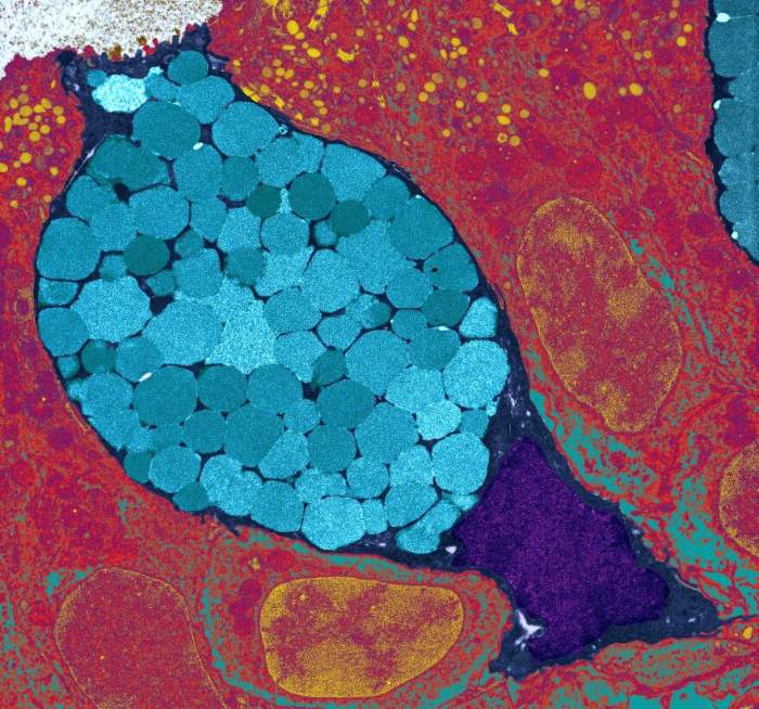
    A nasal weapon: secretion globules

    secretion globules
    Goblet cells. Mucous membranes that are allowed to cool down create fewer “decoys” for cold viruses. (Credit: University of Edinburgh).

    Okay, but what causes a nose to get cold? That’s precisely what Dr. Di Huang and his team at Northeastern University found. Their work focused on small, membrane-enveloped secretory globules that human nasal mucosa produces in reaction to microorganisms, which they reported in 2018. When our immune system detects pathogens, cells in the nasal mucus’ frontal lobe release large numbers of these antibacterial protein-laden vesicles.

    It remained unknown, however, whether or not these vesicles were also created during viral infections, or what they were made of to combat viruses in nasal mucus. Two types of rhinoviruses and a cold coronavirus were used to infect human nasal mucosal tissue samples by Huang and his colleagues.

    Decoys against viral infection

    The experiments revealed that the cold viruses also stimulate the release of extracellular vesicles. However, this happens through a different signaling mechanism than in bacteria, the researchers observed. As a consequence, the membrane-enveloped secretion globules also have a distinct composition: they carry on their surface exactly the receptors that the cold viruses ordinarily employ to hook onto the mucosal cells.

    That is to say, these vesicles serve as a distraction for our nose. These “host” cells really attract viruses, much as a decoy on a military plane might attract a stray missile. These viruses bind to receptors on secretory globules, where they are ensnared and neutralized. According to Huang, the more of these decoys our nose generates, the greater the possibility that viruses will be trapped by them before they can infect the mucosal cells.

    Inhibition of vesicle formation by cold

    Here’s where the cold comes in. To see how much of an impact cold air has on this newly found defensive mechanism, researchers chilled nasal mucosa cultures by five degrees, which is about the same as the amount our noses cool when we step outdoors into the four- to five-degree air. Actually, it came out that the amount of protective vesicles secreted by the nasal mucosa decreased by 42%, and there were less antiviral proteins on the surface of these vesicles as a result.

    As a consequence, we can now explain the cyclical nature of upper respiratory tract infections using a mechanical model. This is due to the fact that our nasal mucosa’s extracellular defensive tactics are weakened by the cold weather, in addition to the fact that cold weather promotes the spread of cold viruses.

    In the fight against the flu

    The scientists have found a novel immunological mechanism in the nose and revealed what hinders this protection. The challenge now is figuring out how to use this natural defense mechanism to lessen the severity of common colds. Nasal sprays that trigger the production of these antiviral vesicles might be a first step.

    A warm nose and a healthy immune system are our main priorities until then. So far, no effective cure for the common cold has been discovered, despite scientists’ best efforts. Creating a vaccine, for instance, is challenging due to the rapid evolution of some viruses, such as rhinoviruses. Similarly, several potential preventative measures have not advanced beyond the laboratory. Studies have demonstrated that zinc supplementation is the only way to reduce cold symptoms and minimize their duration.

  • Viruses and cancer: Discovery of an “impossible” relationship

    Cancer is a terrifying pandemic that hangs over our heads like the sword of Damocles. Innumerable individuals are afflicted by this multifaceted illness, and tens of thousands more must live with the diagnosis every day. Even with the best diagnostic tools and treatment options available, many people still die from it. Doctors are working tirelessly to determine what has triggered this cancerous multiplication of cells.

    Some of the possible causes of cancer have been identified as exposure to toxins or radiation or a family history of the illness, although unlike with many infectious diseases, a direct correlation between cause and effect is usually not sufficient for cancer. The catastrophic transition from a healthy body cell to a tumorous cancer cell is often set in motion by the confluence of a number of elements. However, scientists have barely scratched the surface of this interaction’s intricate complexity.

    Tumor viruses are one aspect that has gained the attention of scientists. The earliest signs that viruses may cause cancer were identified by scientists around the turn of the past century, but this information was largely disregarded for decades. In the past, there was no link between infectious agents and tumor diseases.

    Despite the fact that the link between cancer and viruses was finally proven, scientists in this area faced an unprecedented wall of opposition. Ignorance, but also tenacity, has a long and illustrious history that continues up to the present day. As such, it served as a near-perfect illustration of the typical sequence of scientific progress described by Austrian engineer and author Robert Musil: “Every few years that something till then held to be in error suddenly revolutionizes the field, or that some dim and disdained idea becomes the ruler of a new realm of thought.”

    More and more viruses are being added to the list of “suspects” by cancer researchers, and roughly a dozen have been recognized beyond question, demonstrating the “new realm’s” visible growth. The human papillomavirus and various herpes viruses are two examples of these ubiquitous yet purportedly innocuous infections. Viruses’ causal pathways in cancer are very vaguely understood, but researchers are working to fill up the gaps.

    The discovery of the cancer virus

    Chickens to revolutionize science

    In the year 1909, a farmer pays a visit to a young physician and biologist Peyton Rous at his new job at New York’s Rockefeller Institute for Medical Research. He is concealing a jar with a cancerous chicken under his arm. A sarcoma, a huge cancerous tumor of connective tissue, had developed on the right side of the breast of this striped Plymouth Rock chicken.

    At the time, research on cancer was still in its infancy, but Rous, who worked on the subject, jumped at the chance to do a battery of experiments using the chick and its tumor. So, he decided to produce an extract from the diseased tissue, filter it to remove any remaining cells, and then inject it into young chickens of the same breed. Unusually for the period, the healthy animals also got cancerous tumors of the same sort as a consequence of this experiment.

    With this in mind, Rous deduced that the tumor was caused by a microscopic pathogen present in the extract. He suspected a parasitic creature, probably one of the recently identified viruses. Rous attempted to prove his hypothesis by successfully transmitting additional chicken cancers via this route. His findings and theorization of a cancer-causing virus were published in 1910, setting off a real “tumor wave.”

    Standing on the sidelines

    However, no success was found in using this method to induce growth in any other animal species. Soon after, Rous was marginalized in the scientific community, and his viral tumors were deemed to be a chicken-specific anomaly. In his acceptance address for the Nobel Prize in 1966, Rous recalled, “The results of the sarcoma virus were met with absolute disbelief.” However, “spontaneous” chicken tumors with different morphologies were successfully transplanted and a virus was found in each of them not long after.

    In the 1930s, Richard Shope, a colleague of Rous’s, obtained comparable results using an extract of a benign skin tumor, a papilloma, from the skin of a wild rabbit species. However, these findings were disregarded since they were not thought to be relevant to cancer.

    “Milk factor” instead of viruses

    The standard medical view, that cancer was at most related to a person’s genes or their environment and could not be caused by viruses or other pathogens, appeared to be impervious to new evidence. Under the banner of “What must not be, cannot be,” any and all theories involving viruses that cause cancer were dismissed as ridiculous. In the 1930s, the United States’ highest health authority convened a council of specialists who issued a formal guiding principle that viruses and other organisms could be ruled out as causes of cancer.

    Due to the atmosphere, few scientists were willing to publicly provide proof of the presence of cancer viruses. Breast cancer-causing viruses were first identified in mice by John Bittner, a researcher at the leading cancer research facility in the United States, in the late 1930s. Instead of calling the cancerous agent a virus in his paper, he called it a “milk factor,” neutrally avoiding the risk of losing his job and future research funding.

    The breakthrough

    The significant change, and the subsequent rise of cancer virus researchers, did not come until the late 1950s. There were two primary causes that resulted in this change: Scientists found that some viruses could introduce their genetic material into the host cell’s DNA. This modified the cells irreversibly without killing them. However, numerous scientists started to find tumor viruses in their lab tests, making it impossible to deny the undesirable reality.

    According to a long-held theory, cancer and infections produced by viruses or other organisms could not be linked to one another. But a decade later, in 1966, Peyton Rous was honored for his foresight and tenacity with the Nobel Prize in Medicine, and the virus he discovered was given the honorific name “Rous sarcoma virus.“

    Looking for the virus-cancer mechanism

    Genome thieves

    Cancer cells are the progenitors of more cancer cells and pass on their destructive capacity to their children, making them the ultimate misfits. Yet they originate in healthy tissue. But, how? What part do viruses and other pathogens play in this process? Caltech cancer researcher Renato Dulbecco was pondering this very subject. As a result of Peyton Rous’s rehabilitation, study into tumor viruses is no longer taboo, although still very little is known about how they work.

    In 1960, he and his colleague Marguerite Vogt undertook laboratory experiments in which they infected hamster cells with a polyoma virus, a DNA virus with little complexity. The cells started to transform as expected, and tumors started to form. However, they immediately noticed something peculiar: the viral reproduction ceased along with the transition to cancer cells. Their numbers did not increase or decrease as they would during an actual infection since they were not able to exhaust the cell’s resources on viral production. Sure, but why?

    The virus always leaves behind traces

    Dulbecco could only think of two possibilities: either the virus got killed out in the process of the cell’s metamorphosis, or it survived the transformation but was unable to multiply. After much effort and testing, the scientist finally got an answer. He discovered the virus’s “footprints” in the changed cells, disproving the original hypothesis and confirming the infection was still there.

    Using molecular biology techniques, he was also able to show that the virus was inactive since a key piece of its DNA had been incorporated into the host cell’s genome, rendering it inaccessible for viral replication. Only around seven proteins were encoded by the viral DNA that was added, but that seemed to be enough for cell processes to go haywire.

    Contrary to dogma

    A young scientist at the University of Wisconsin named Howard Temin had less luck than Dulbecco did when he published his findings and received both praise and criticism for them. Unlike Dulbecco, Temin studied an RNA virus (the Rous sarcoma virus) in his tests; yet, he reached the same conclusion as Dulbecco in a 1964 paper: the virus must add something to the cell’s genome that initiates the cell alterations. According to him, before inserting its genetic material into cellular DNA, the virus was “translating” its RNA genome into DNA, called the “provirus.”

    However, he made a huge “error” by proposing this theory, since it challenged the prevailing paradigm in molecular biology at the time. This dogma states that information in live cells always flows from DNA to RNA and then to protein, but never the other way around. For this reason, most of Temin’s scientific peers would have considered his concept of a provirus that employed this “wrong” path to be heretical. The response was just as furious and condescending.

    Again, it took almost a decade and the “administrative aid” of a coworker before the previously disgraced scientist was vindicated. Temin and the biologist David Baltimore delivered the killing blow to the doctrine at the decade’s close in the 1970s. Both separately found an enzyme in tumor virus-infected cells that can convert RNA to DNA. There was experimental proof that this “provirus DNA” was integrated into the cellular genome.

    The seminal work on the biology of tumor viruses by Dulbecco, Temin, and Baltimore was recognized with the Nobel Prize in 1975. However, the three of them were unable to resolve the lingering mysteries of how viruses cause cancer and whether or not they also cause cancer in humans. “I do not believe that infectious viruses are the causes of most human tumors, but I think that viruses can provide us with models for the development of cancer,” Temin said cautiously in his Nobel Prize acceptance address.

    Looking for a cancer gene

    Can cancer viruses replicate without causing disease?

    Two researchers at the U.S. National Cancer Institute, Robert Huebner and George Todaro, experimented with the Rous virus on various mammals in the 1970s, shocking the scientific community just as they were coming to terms with the discovery that cancer viruses, including and especially RNA viruses, incorporated part of their genetic information into the DNA of the cell. They found, to their shock, that viral DNA could be detected in tumor cells even when the tumors had not been caused by the virus but rather by physical or chemical effects. How could this be explained?

    The “virogen-oncogene hypothesis”

    Huebner and Todaro proposed that viral DNA doesn’t come “out of nowhere” since cells must already have viral genes embedded in their genetic material, which were incorporated into the genome at some point during evolution. These conserved DNA regions are inactive under normal conditions but become functional when exposed to certain stimuli, allowing them to produce viral proteins and, in turn, new viruses. However, they postulated that some of these “virogens” also operated as “oncogenes,” causing the development of tumors as well.

    Hunting down the tumor gene

    If this hypothesis were to hold, then the Rous oncogene must also be present in the cellular DNA of normally developing animals. Michael Bishop and Harold Varmus of the University of California, San Francisco, began a mission to match the genome of the Rous sarcoma virus with chicken DNA for this very aim. But this would be very difficult to do without high-tech equipment like PCR or sequencing devices.

    Fortunately, in 1971, a serendipitous discovery by Peter Vogt of the University of Southern California revealed a viral mutant that was capable of replication but seemed to lack the potential to produce malignant tumors. The virus’s genome was 15 percent shorter than average, suggesting that this missing part could include the critical tumor gene(s).

    This was the key piece of information that helped Bishop and Varmus solve the case. This allowed them to determine whether or not the “src” gene fragment was present in the cellular DNA of their guinea pigs and, if so, whether or not it functioned as a possible cancer gene. However, scientists were surprised to discover that the virus was present not just in chickens, the primary hosts of the Rous virus, but in birds of every genus and subgenus.

    But that wasn’t the end of it; other researchers soon identified src variations in guinea pigs, rabbits, rats, snakes, and even humans. The oncogene postulated by Huebner and Todaro seemed to be found. Bishop and Varmus, though, were not confident in the claims. They were wary about its extraordinarily widespread distribution. The origin of the gene still remained unknown; did this gene really come from the Rous virus?

    Oncogenes: a biological enigma

    The enemy within

    An essential step forward accomplished with the discovery of a cellular proto-oncogene. Not much time had passed, yet scientists had already uncovered over 40 distinct retroviral oncogenes. They speculated that the majority of cells had gene sequences that were homologous to a viral gene known to cause tumors. Tumor viruses and other external factors could contribute to tumor development, but there was also an “internal adversary” that might be just as crucial.

    However, more questions were created by the new data than they answered. Among the most crucial, in Varmus’s words, “Did the evolutionary conservation of the src gene indicate its cellular origin? Or could it still be a virus-derived gene that was just better conserved than comparable viral elements?”

    Own cell instead of being smuggled in

    Rapid responses to this question emerged from many different fields. Sequence analysis, however, provided the clinching evidence. It demonstrated that the src gene was broken up into many segments by introns (“junk DNA”), which were typical of biological DNA but were rarely seen in viral genes.

    Harold Varmus reflected on this, saying, “All our arguments and experiments led to only one conclusion: the precursor to src was a conserved – and therefore vital – cellular gene that found its way into the Rous sarcoma virus by recombination.” So, it seemed that the proposed evolutionary route of the “virogen oncogene” concept must have followed an exactly opposite direction.

    Since the oncogene’s precursors have been conserved throughout evolution, this explained why they are found in so many different types of cells. “Far from being a harmful element just waiting to be activated by a carcinogenic signal, the precursors of viral oncogenes seem to have an impact on the organism to have an important function, otherwise they would not have been preserved in the course of evolution,” said Varmus. These genes are, in fact, involved in essential cellular processes, including multiplication and differentiation.

    Hostile takeover

    How, therefore, could this normally helpful gene cause an infected cell to go berserk? Bishop and Varmus hypothesized that the retrovirus identifies the benign proto-oncogene during infection and alters it into the malignant oncogene. During this process, known as transduction, the virus inhibits the gene’s regular function and removes it from the organism’s regulatory system. Therefore, the misplaced and overactive genes have undesirable effects, such as excessive cell multiplication.

    The two scientists speculated that Huebner and Todaro’s peculiar results might be explained if the transition was triggered by chemical or physical forces. In 1983, researchers found a group of cellular genes that were structurally similar to viral cancer genes and oncogenes from non-viral cancers, lending credence to this theory.

    Viruses as a possible cause of human cancer

    Initial evidence for human T-virus type 1

    By pinpointing the “inner enemy” and showing that viruses might play a role as triggers in carcinogenesis, Bishop and Varmus have thrown open the floodgates for contemporary cancer research. They shared the Nobel Prize in Medicine in 1989.

    However, they encountered resistance and even contempt for their ideas. Their hypothesis, that a comparable process could possibly contribute to human tumor formation, was at odds with the current scientific consensus. But this perspective held that although viruses were identified as a cause of tumor disorders in animals, there was less evidence to support a similar link in humans. But they weren’t completely wrong since hereditary or environmental factors are still considered to be the main causes of cancer in humans.

    The first human retrovirus

    However, this started to change in the early 1980s. In 1980, a patient with cutaneous T-cell lymphoma, a benign skin ulcer, was investigated by virus researcher Robert Gallo and his colleagues at the National Cancer Institute in the United States. He analyzed the tissue samples and found a retrovirus, a kind of virus that had never been seen in humans. While these RNA viruses, to which the Rous sarcoma virus also belongs, had been studied for quite some time in animals, this was the first time that they had been found in people.

    Gallo and his Japanese colleagues continued studying its biology and ecology in the years that followed. Scientists found an unexpected link between the virus and adult T-cell leukemia lymphoma; whereas most infected people had relatively mild skin changes over time, a small percentage had acquired this aggressive type of leukemia.

    This kind of cancer often does not manifest until the age of 60, but once it does, it generally results in a swift death. It is estimated that just 1–4% of infected people actually acquire leukemia, but all patients carry the virus anyway.

    And the Leukemia

    So, was the virus the cause of cancer? There had to be more evidence than just finding a virus in a cancer cell. But in 1982, Gallo and his group found the viral “culprit” in the virus’s DNA. Since there was no homolog of the viral gene “tax” in the human cell genome, “tax” could not be considered an oncogene in the traditional sense. But when it was inserted into the cell’s DNA, it activated particular T cell growth factors, leading to the unchecked multiplication characteristic of leukemia. The specifics of this mechanism were, however, still little known.

    The pathogen, now known as HTLV-1, or human T-cell leukemia virus, made medical history as the first virus known to cause cancer in humans. This discovery sparked what has become a decades-long effort to identify and characterize additional viruses that might cause human tumors. Even while the hepatitis C pathogen was now thought to have a role in certain kinds of liver cancer and maybe even in some forms of non-lymphoma, Hodgkin’s other RNA viruses had thus far been on the suspect list at best, with nothing yet confirmed.

    Human papillomavirus (HPV) and cervical cancer

    Forming a model

    Human papillomavirus (HPV) is a tiny, contagious virus that may manifest in anyone’s cells. The current global prevalence of this DNA viral infection is estimated to be above 25 million. The sexually transmitted disease affects almost half of the male and female population.

    Infected people may not even realize they have it. The virus is asymptomatic and may live in the skin and mucous membranes for a long time; in rare instances, benign warts may appear. In addition to its pervasiveness, HPV is also remarkably diverse, with over 130 distinct strains having been found to date. And 18 of them are particularly worrisome since they belong to high-risk categories and may cause cancer. Discovered in 1983, this link between a virus and cancer is now one of the best studied and proven anywhere. Some virologists think that HPV is responsible for at least 600,000 instances of cancer globally; other estimates place the number at between 12 and 15 percent of all tumor cases.

    Two proteins that sneak into cells

    The human papillomavirus types 16, and 18, are mostly to blame. More than 90% of head and neck cancers and 99% of cervical cancers have these abnormalities. In most cases, the human immune system can clear up an HPV infection in around ten months. Changes occur, however, when risk factors are introduced, such as smoking, an impaired immune system, or a particularly aggressive viral strain:

    Two viral proteins, E6 and E7, are produced by the infected cells. When bound to a particular cell protein, E6 triggers that protein to label the tumor suppressor gene P53 for destruction. Consequently, the cell is deficient in a key component for preventing mutations and cancerous transformations. Another viral protein, E7, is responsible for cell death by “hijacking” a cellular protein engaged in crucial regulatory mechanisms. This causes the release of transcription factors that actively promote cell multiplication. Two proteins assist the virus in disrupting the cell’s basic metabolic and developmental processes, leaving the cell vulnerable to mutations.

    However, this is by no means the sole mechanism by which HPV causes cervical cancer. In fact, new mechanisms are disclosed virtually every week. These two, though, are the sharpest and most consequential.

    The dirty dozen

    Researchers have had a hard time finding new tumor-inducing viruses due to the intricacy and variety of the processes and regulatory systems already engaged. This is one of the reasons why just a handful of anti-cancer drugs that target viruses have been shown to be effective in people.

    Epstein-Barr virus (EBV) for nasopharyngeal cancer and Hodgkin’s lymphoma; hepatitis viruses B and C for liver cancer; human T-cell lymphotropic virus type 1 (HTLV-1) for cervical cancer; and human herpesvirus 8 (HHV-8) as a trigger for Kaposi’s sarcoma are all well-established viruses that cause cancer in humans. Polyomavirus and hepatitis C for non-Hodgkin’s lymphoma, and an unidentified agent for a kind of pediatric leukemia, remain on the list of possible tumor-causing viruses. The tally of cancer viruses in humans that have been “condemned” continues to increase today.

    Helicobacter pylori and stomach cancer

    Several kinds of cancer in humans now have a known link to infections with certain viruses, bacteria, and parasites. 15–20% of all malignancies are believed to have an infectious cause. But we weren’t always aware of the cancer-causing bacteria. In fact, this medical dogma was toppled anew by a dogged scientist, a little bit of self-experimentation, and a giant leap forward. The Australian scientist Barry Marshall, his colleague Robin Warren, and the rod-shaped bacteria Helicobacter pylori are the story’s primary characters.

    In 1982, the medical community accepted the dictum that “Bacteria are everywhere—but the human stomach is bacteria-free.” Because of the very acidic conditions present in the stomach, it was often believed that no bacteria could thrive there. And that was not all. The majority of medical professionals and scientists in the field of pharmacology, as well as patients, attributed the pain of gastric mucosal inflammation and subsequent gastric ulcers to the presence of acid in the stomach. The pharmaceutical industry promoted acid blockers of different sorts with the slogan “No acid, no ulcer,” and sales were booming as a result.

    Its home is the stomach

    Pathologist Robin Warren found something that shouldn’t be there when he examined tissue samples from stomach ulcers and irritated stomach mucosa under a microscope: bacteria that are full of mucus. And that was the case in over half of the samples he tested. The number of these cells seemed to be correlated with the degree of inflammation. Warren consulted with doctor Barry Marshall after concluding that his finding must be significant.

    As a team, they achieved the first successful cultivation of the bacteria. They could now undertake microbiological experiments to learn how the bacteria manage to thrive despite the harsh circumstances of the stomach. To begin attaching to the mucosal cells, bacteria first use their flagella to dig deeply through the protective mucus layer. Meanwhile, they used an enzyme called urease to convert the uric acid produced by the cells into carbon dioxide and ammonia. This created a neutral environment that bypassed the stomach’s defense against microbes.

    A sip alters the course of history

    However, these results did not establish beyond a reasonable doubt that this particular bacteria caused an inflammation or cancerous tumor. Because the conclusive evidence was still absent. In July of 1984, after months of testing, Marshall decided to take a daring step: he began testing on himself. On the informal principle of “a small sip for a person-a big sip for medicine,” he consumed a brackish solution containing billions of germs. As Marshall reflected, “Everyone said, you’re crazy. It seemed like too incredible an explanation for something as complex as stomach ulcers.”

    In a matter of days, he started feeling sick, like he had the flu, and in two weeks, he had gastritis. With the use of a tissue sample, he was able to see that his mucosal cells were covered in a thick layer of germs. Thus, Marshall successfully exposed Helicobacter as the true culprit in the inflammatory process. At that time, though, the link he suggested between the bacteria and stomach cancer had not been proven.

    The murderer’s in the blood

    However, since then, his theory has been independently verified: antibodies may indeed identify the bacteria in the blood. Three further significant epidemiological investigations followed this. Scientists looked for Helicobacter antibodies in the blood of thousands of stomach cancer patients, whose blood was collected and preserved decades ago as part of earlier research, and compared the findings to the values of a similarly large sample of patients who did not have cancer of the stomach.

    Those who were infected with Helicobacter 20 years ago had a six-fold increased risk of developing cancer compared to those who were now free of Helicobacter. These findings were so compelling that in 1994, the World Health Organization’s International Agency for Research on Cancer (IARC) officially labeled Helicobacter pylori a class 1 carcinogen, the first identified bacteria capable of causing cancer.

    Terms related to viruses, cancer, and genes

    Most of what we know about how genes play a role in cancer was uncovered via studies of tumor viruses. The relative simplicity of the viral genome has been the only thing that has allowed researchers to identify, or at least narrow down the DNA building blocks involved in the formation of human cancer.

    The link between viruses and cancer has been recognized to exist in animals since the middle of the past century, but only in people since the 1980s. Unlike animal tumor viruses, however, the link between human cancer viruses and the disease is based on correlations or presumed processes rather than a clear cause-and-effect premise. The viruses have a role as either cofactors or cocarcinogens in this scenario. This demonstrates undeniably that the process of carcinogenesis in humans is intricate and multi-staged.

    At least six families of DNA viruses and two families of RNA viruses are suspected in the development of cancerous human tumors. Among the examples are Retroviridae and Flaviviridae as RNA virus families and Hepadnaviridae, Herpesviridae, and Papillomaviridae as DNA virus families. 

    RNA viruses:

    Those are the viruses that employ RNA rather than DNA to store their genetic information. Single and double-stranded forms of the RNA are possible. Examples of RNA viruses include influenza, polio, and Ebola; the hepatitis C virus has been linked to cancer.

    Retroviruses:

    The retrovirus family is also an RNA virus. To integrate their genome into the host cell’s chromosome, retroviruses employ the enzyme reverse transcriptase to create a DNA translation of their genome. Here, it is incorporated into the DNA of the cell for good. The AIDS virus and HTLV-1, a virus linked to leukemia, are examples of such pathogens.

    DNA viruses:

    These viruses have their genetic material in the form of DNA and may thus integrate it into the host cell’s genome without first requiring “translation.” Herpes viruses, human papillomavirus (HPV), linked to cervical cancer, and Epstein-Barr virus, linked to certain types of leukemia, are all examples.

    Transduction/transformation:

    This is the process through which healthy cells mutate into cancerous ones. This mechanism has been greatly elucidated by the capacity of viruses to convert cells in culture. The altered cells display hallmarks of cancerous tumors, including unchecked multiplication, reduced reliance on supporting tissue, and less need for external growth hormones. When transplanted into a new host organism, some altered cells may potentially become cancerous.

    Tumor virus transformation of a cell always exhibits the following three characteristics: it is a “one-hit” process, meaning that the transformation is triggered by a single viral infection of the cell. In addition, the cell retains some viral genetic information, and the transformation is maintained by the continued production of a subset of viral genes.

    Oncogene:

    A gene that has a significant role in causing or facilitating the progression of cancer. Point mutations, rearrangement of its DNA building blocks, loss of individual segments, and excessive, uncontrolled gene expression triggered by viruses all contribute to its activation and transformation into a tumor gene. The Greek term for “mass” or “lump” is whence we get our modern word “onco.”

    Cellular Oncogenes:

    An oncogene may mean either a normal gene that has been changed to become active in tumor cells or a cellular gene that has been activated in tumor cells.

    Viral Oncogenes:

    DNA viruses may cause cancer because they include a gene that isn’t found in cells but has the potential to do so when infected, or because they contain a version of a normal cellular gene that has been changed or activated to operate as a tumor inducer.

    Proto-oncogene:

    A precursor of both viral and cellular oncogenes, a normal gene of the cell that may be turned into an active oncogene by external factors.

    Tumor suppressor gene (TSG):

    Genes encoded by a cell’s own DNA inhibit cancerous transformation. These genes may have a bigger effect on carcinogenesis than the active oncogenes.


    Bibliography

    1. Viruses Associated with Human Cancer – PMC (nih.gov)
    2. Zur Hausen H. “Novel human polyomaviruses – re-emergence of a well known virus family as possible human carcinogens”. International Journal of Cancer.
    3. Woolhouse M, Scott F, Hudson Z, Howey R, Chase-Topping M (2012). “Human viruses: discovery and emergence”. Philosophical Transactions of the Royal Society of London. Series B, Biological Sciences.
    4. Koonin EV, Senkevich TG, Dolja VV. “The ancient Virus World and evolution of cells”. Biology Direct. doi:10.1186/1745-6150-1-29.
    5. “Virus Taxonomy: 2021 Release”. talk.ictvonline.org. International Committee on Taxonomy of Viruses.
    6. Breitbart M, Rohwer F. “Here a virus, there a virus, everywhere the same virus?”. Trends in Microbiology. doi:10.1016/j.tim.2005.04.003.
  • Virus: One of the most successful models of evolution

    Viruses are an excellent example of successful evolutionary adaptation since they outnumber all living cells, are present in all living things, and have been there since the beginning of life. But why are these parasitic cellular organisms so effective? And why do coronaviruses, Ebola, and influenza, all of which are enclosed RNA viruses, cause epidemics time and time again? It’s undeniable that viruses are quite effective in hijacking cells. They are biologically and structurally suited to infecting cells and turning them into replicating viral factories. Viruses have evolved an incredible range of tactics, each tailored to a certain host. However, it is plausible that a virus-like structure existed from the origin of all life, and that the cell parasitism of viruses was a key factor in the evolutionary breakthroughs that followed.

    The first “virus”: Tobacco mosaic was originally attributed to what Beijerinck, in 1898, was the first to identify as a “virus“. He demonstrated that the inciting agent moved about in agar gel, making it clear that it was a ‘contagium vivum fluidum,’ or a soluble infectious agent, and not a ‘contagium fixum,’ like a bacterium.

    Viruses are in every ecosystem, every living being

    Viruses are the most widespread form of life on Earth, and they may be discovered in almost every environment. There may be ten to one hundred times as many viruses as there are live cells. However, just a tiny proportion of the viral variety has been discovered thus far.

    Viral roommates

    phages
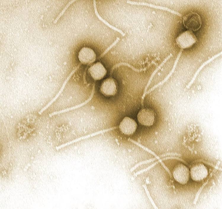
    The vast majority of viruses in and on our bodies are phages, which infect the bacteria that live on us. Credit: Vincent Fischetti, Raymond Schuch / PLoS Biol 2/2/2004: e53 CC-by-sa 2.5

    This includes habitats that are quite near to us, such as the air we breathe and the skin we have. After conducting a “dragnet search” for viral genes, scientists uncovered hundreds of millions of sequences. Around 90% of them have never been studied by scientists. However, the findings demonstrated that, like the bacterial microbiome, each region of the body has its own distinct population of viruses. And there are still viruses that haven’t been discovered that can infect our gut. Studies uncovered one of these pathogens, which affects the bacteria that live in the human intestine and goes by the name Escherichia coli.

    Of course, we aren’t the hosts for the great majority of the viruses that live on and in our bodies; they are peaceful inhabitants. These viruses prefer to infect bacteria, which they then employ as a host for reproduction. Initial results suggest we may harbor millions more of these bacteriophages. The viruses that may really make us ill are a tiny fraction of this massive population. However, as the current Corona epidemic shows, the effects of these events on human health are far more severe.

    Hosts throughout the family tree of life

    virus life tree
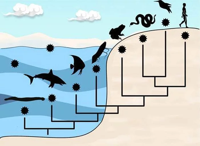
    Almost every group of living things may be infected by viruses. ©University of Sydney

    The host range of viruses, however, covers the whole tree of life, not just humans. Viruses may cause harm to almost every living thing. This diversity includes everything from nucleus-less archaea and bacteria to eukaryotic unicellular animals like insects and other invertebrates, all the way up to vertebrates like fish and humans. Aside from viruses that only infect algae, practically all higher plants have their own. At least in part, the tobacco mosaic virus, which causes illness in tobacco plants, was the first virus whose make up scientists were able to understand in 1935.

    Nonetheless, most virus species are highly specialized, meaning they can infect just one host or a small group of closely related animals. Because they solely go after our resident bacteria, the millions of bacteriophages we harbor pose no threat to us. We are also immune to viruses that only attack plants and other invertebrates. Such barriers appear conceivable since creatures that are evolutionarily far away also vary in considerable detail in their cell biology. Since these conditions are so dissimilar, it’s challenging for a virus to reproduce in any of them.

    From deep rock to the atmosphere

    Viruses have been able to colonize practically every ecosystem on the planet due to the variety of hosts they may infect. They populate the watery environments of the world, from the tiniest puddle to our sewage to the grandeur of the seas, and the soils of practically every ecosystem on land. More than 10 million viruses may be found in just a single drop of saltwater. Viruses may be carried great distances in the air by both sea spray and desert dust.

    As a consequence, more than 800 million viruses per square meter float in the atmospheric boundary layer every day. From a height of 2.5 to 3 kilometers, these airborne pathogens are carried by winds all across the world. It’s quite possible, the scientist continued, for a virus to spread through the air on one continent and then land on another. That’s also why related viruses may be discovered hundreds of kilometers away.

    Viruses are ubiquitous, and may be found in even the harshest settings on Earth. Such places include Antarctica’s subglacial lakes, the hot springs and hydrothermal vents of the deep sea, and the rock strata at a depth of more than a kilometer below the ocean bottom. Based on sampling, in the deep biosphere, they may even constitute the majority of the biomass.

    Viruses are ubiquitous, which demonstrates how extensive the virosphere, or viral universe, really is.

    That begs the question, however, what exactly is their magic formula?

    What are viruses?

    Maximum efficiency in a minimal design

    Viruses are, at their most fundamental level, just a bunch of DNA or RNA enclosed in a protein coat. In the natural world, they are the minimalists. Their basic structure is like a doghouse compared to the Sistine Chapel when a live cell is being considered. Nonetheless, this does not make viruses any less effective.

    Highly variable genome

    viruses
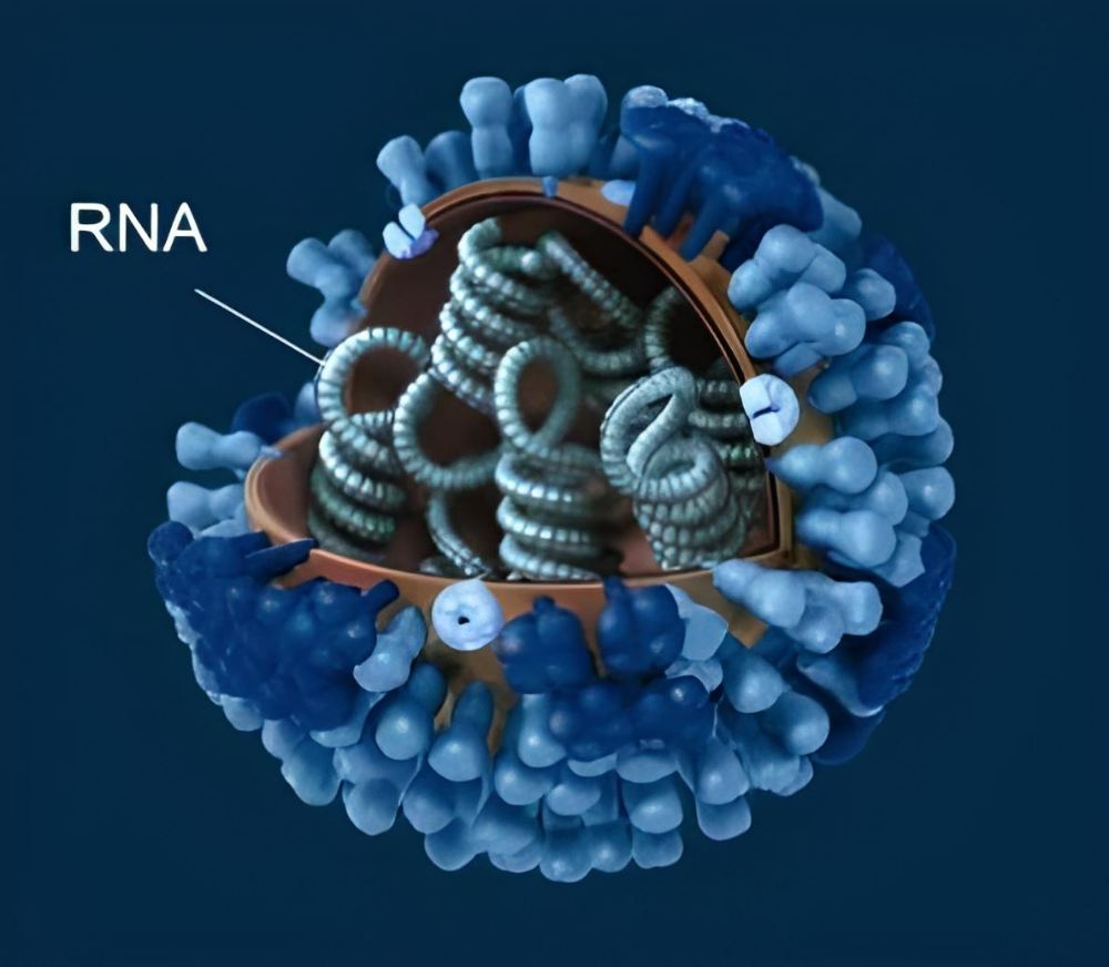
    DNA is replaced by RNA in influenza viruses. © CDC

    Viruses have evolved to fulfill their specific role in the world, and they carry with them all the tools necessary to maintain their species via reproduction. The viral genome encodes the proteins that make up the virus’s envelope and other critical enzymes. Genome size varies widely amongst different viral families, from tiny bacteriophages with just approximately 3,500 bases, to large polioviruses with 7,500 bases, to enormous viruses with 1.2 million bases.

    Although all living things pass on their genetic information through the hereditary molecule DNA, the genetic material of viruses is very diverse. Depending on the kind of virus, it may be either DNA or RNA and can have either a single-stranded or double-stranded structure. Herpes viruses, papillomaviruses, and the smallpox virus are all examples of DNA viruses.

    Many pathogens of serious and widespread infectious illnesses are RNA viruses, including the newly discovered coronavirus SARS-CoV-2, Ebola, measles, and influenza. Furthermore, many illnesses spread by mosquitoes or ticks, such as dengue, Zika, West Nile fever, and yellow fever, are caused by RNA viruses. The HIV virus, responsible for AIDS, likewise uses RNA as its genetic material.

    Effectively built armored capsule

    The viral capsule, or capsid, is the second part of every virus. It contains many copies of a single or small number of proteins and encloses the viral DNA. This results in a compact full capsid assembly that takes up little room in the viral DNA. However, viruses with just their capsid as an exterior shell are less adaptable because of their efficient manufacturing, which prevents the protein composition of the capsid from changing readily.

    poliovirus 2
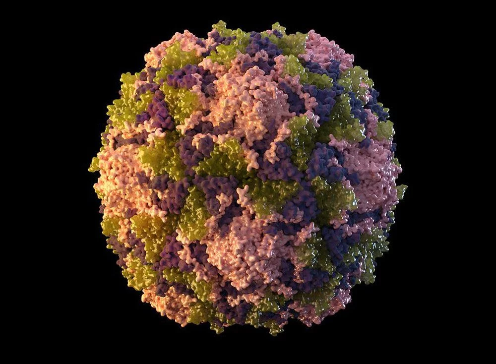
    To create its capsid, the poliovirus replicates each of its four proteins 60 times. © CDC/Sarah Poser

    The capsid’s protein arrangement is another feat of natural efficiency: The proteins arrange themselves into a pattern of recurrent, identical basic units, which ultimately coalesce into the symmetrical viral capsid. Proteins may be thought of as disks that assemble into a helix, enclosing the viral DNA. The tobacco mosaic virus, the influenza virus, the measles virus, and the rabies virus are all examples of viruses possessing a helical capsid.

    The icosahedron is another frequent capsid design; it has 20 sides and is built from basic units that are triangles composed of three copies of the same protein. The icosahedron has 20 of these triple units or multiples thereof, depending on the virus. Small and basic, its capsid is shared by the rhinoviruses and polioviruses that cause the majority of the colds in a country. They have a capsid made up of four distinct proteins, and each of those proteins creates 20 of these simple triple units.

    Stolen envelope

    influenza virus
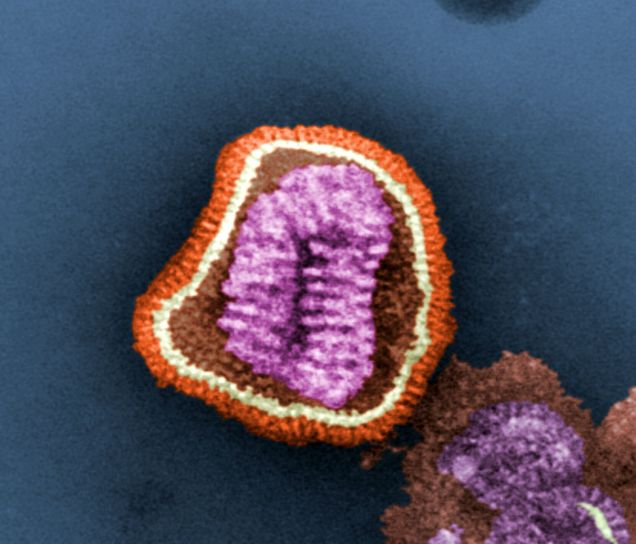
    The influenza virus, like other viruses, has an outer membrane called the viral envelope. This membrane is made of lipids and contains envelope proteins. ©CDC

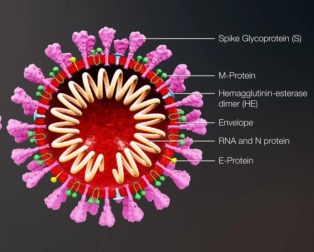
    In addition to their DNA and capsid, many epidemic-causing viruses, such as the severe acute respiratory syndrome coronavirus 2 (SARS-CoV-2), also feature a viral envelope. It is similar to the cellular membrane in that it is a double lipid membrane with embedded proteins. The nucleocapsid is protected from the environment by this envelope, which completely encloses the virus.

    Most enveloped viruses obtain the viral envelope by budding through the cell membrane and enclosing a portion of the host cell membrane around the virus, together with viral proteins already generated by the host cell. In other words, viruses may skip making their own blueprints for the double lipid membrane and instead just utilize the envelope. The membrane envelope facilitates the entry of these viruses because their envelope may readily fuse back to the cell membrane, releasing the mutant-filled capsid into the inside.

    Why can enveloped viruses spread so easily?

    Epidemics are often caused by enveloped viruses, such as the coronavirus SARS-CoV-2, influenza, smallpox, and Ebola. The viral envelope facilitates the ability of these viruses to alter their behavior in response to environmental changes, increasing their chances of infecting people.

    Advanced camouflage

    One reason is that many viruses have evolved a “camouflage coat,” in which the membrane of the virus’s coat and some of the coat proteins found inside it closely resemble those of common host molecules and cell components. This aids the pathogen’s efforts to hide from the immune defense system, increasing the likelihood that it will go undetected at first. Some coronaviruses, for instance, contain envelope proteins that are structurally similar to some human immunoglobulin G antibodies.

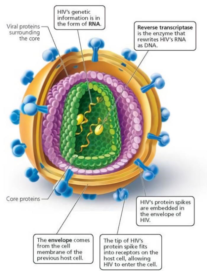
    Many different viruses use sugar molecules produced inside the body as a disguise. Glycans, which are sugar molecules, bind to the proteins of the envelope and hide their terminal amino acids. Moreover, the virus’ sugar coats mimic those of the body’s own proteins, making it more difficult for the immune system to identify it as an invader. These glycans cover almost the entire surface of the HIV virus like a camouflage cloak; they account for over half of the total mass of the envelope protein.

    Deception

    hiv structure 2
    Similarly, the glycoproteins of the HIV virus’s envelope serve as a disguise from the immune system.

    Additionally, viral coat proteins often exhibit protrusions or loop-shaped side chains near their termini. Although they are subject to alteration as a consequence of mutations in the virus’s genome, these components are not essential to the pathogen’s operation. As a result, it is able to rapidly and regularly alter its outer structure. This renders the immune defense antibodies useless since they no longer have any places to recognize or dock with the pathogen.

    The Ebola virus utilizes a more ingenious strategy: it creates “decoys,” or little bits of a membrane with linked protein sections, to lure the host’s antibodies and divert them from the true viruses before they leave the host cell.

    Precision docking

    virus cov 2
    Docking with the host cell is accomplished by the SARS-CoV-2 spike protein on the viral envelope. Credit: GettyImages

    Enveloped viruses may rapidly adapt to new hosts and transcend the species barrier from animals to humans, which is part of the reason why they are so effective at causing new epidemics. Certain viruses, such as influenza and coronaviruses, employ components of an envelope protein to attach to particular receptors on their host cells, while non-enveloped viruses have a more difficult time modifying their capsid.

    The virus can only infect cells if its binding site is properly configured. This also affects which organs or tissues a virus infects, since not all cells have the same receptors. For instance, the SARS-CoV-2 coronavirus’s spike protein is shaped in such a way that it docks with the ACE2 receptor on human respiratory and lung cells. In contrast, the TBE virus that is responsible for tick-borne meningitis has a binding site that is specific to the receptors found on nervous system cells.

    Predestined for the origin of the species

    In any case, the binding site of encapsulated viruses is very malleable and may undergo rapid morphological shifts due to mutations. Because of this, these diseases are able to modify their cell receptors to interact with those of new hosts. Additionally, they are able to communicate with people of various species. Most infectious illnesses that have recently migrated from animals to people are caused by enveloped viruses, and this is not a coincidence.

    Recent examples include SARS-CoV-2, a coronavirus that developed in bats but then underwent a series of modifications that made it transmissible to humans. After infecting another species, perhaps pangolins, it made the leap to people in China in late 2019. There is debate about whether the coronavirus fully adapted to its animal hosts or to its initial human hosts.

    Moreover, influenza viruses are proficient “species barrier jumpers.” Many of the influenza viruses now making the rounds first appeared in birds, and new avian influenza strains are constantly emerging, some of which have begun to show symptoms of adaption to humans. Furthermore, the 1918 influenza pandemic virus also spread from birds to people. In 2009, its offspring were responsible for another pandemic, this time caused by the “swine flu.”

    How do viruses multiply?

    Viruses can only replicate inside living cells. They can only reproduce by taking over the cells of other organisms and using their resources for their own ends. They lack the ability to generate their own genome or envelope and lack the metabolic machinery to manufacture energy-rich molecules like ATP. They must always live within a host cell.

    DNA viruses: taking over the cell machinery

    dna virus
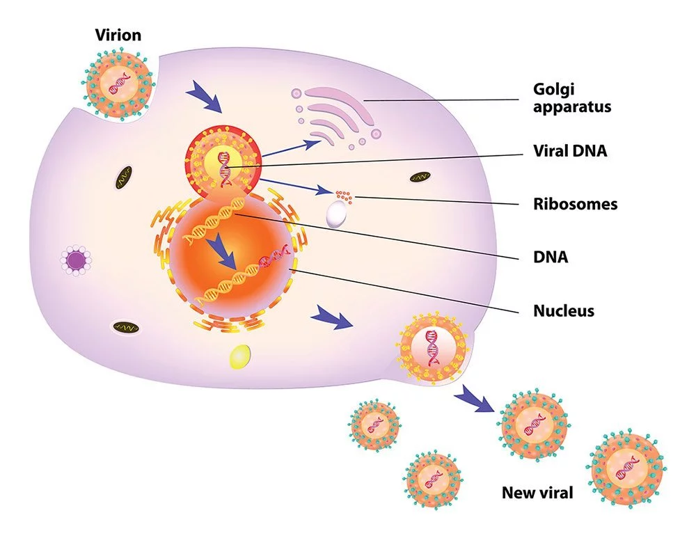
    DNA viruses employ the host cell’s transcription and translation machinery to spread their genetic material within the host cell. ©ttsz/ iStock.com

    When a virus invades a cell, it immediately starts its aggressive takeover. Infectious agents like herpesviruses, papillomaviruses, and others that employ double-stranded DNA to replicate do so by transporting their DNA into the nucleus of a host cell. They trigger the cell’s own enzymes to decode the viral DNA and produce mRNAs. After leaving the nucleus, these mRNAs make their way to the ribosomes, where they are used to make viral enzymes.

    The second phase involves the virus using these enzymes to totally take over the cell and turn it into a viral factory. At the same time as the proteins for the capsid and the viral envelope are being made on the ribosomes in the nucleus, fresh copies of the viral genetic material are being created in rapid succession. During budding, the viral envelope encloses the newly formed virus after it has penetrated the cell membrane and linked the viral genome to the capsids. The aftermath is a dead cell and a flood of new viruses.

    Coronaviruses: multiplication with its own RNA factory

    The coronavirus has a unique method of reproduction. This is due to the fact that instead of DNA, it has a single strand of positive RNA, which has both benefits and drawbacks when it comes to gaining control of the cell’s machinery. The downside is that, since our genome is stored as DNA, our cellular machinery was not developed to duplicate RNA. The only purpose of RNA is to transport genetic information from the nucleus to the ribosomes. The ability to replicate RNA into RNA is not supported. In order to replicate, the coronavirus must first set up its own duplicating machinery within the host cell.

    However, once inside the cell, a portion of the virus’s RNA strand may make its way to the ribosomes, where it can be read without any additional processing. From this, the primary protease is created and used to break down the lengthy protein chain generated by the ribosomes.

    Coronaviruses multiplication with its own RNA factory 1
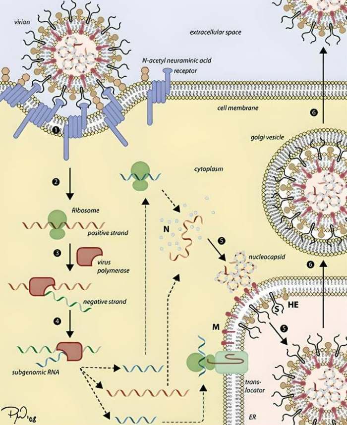
    The cycle of coronavirus replication.

    This generates the mechanisms necessary for replication, turning the cell into a production facility for more coronaviruses. The process begins with the viral enzyme known as polymerase copying the virus’s original RNA strand in order to create a complementary strand. This strand of negative RNA is currently being used as a transcription template to generate an abundance of new, positive RNA strands, which will ultimately act as the viral genomes. At the same time, the ribosomes read a short section of the opposite RNA strand and synthesize the capsid and new viral envelope proteins.

    Copying errors make RNA viruses more adaptable

    In addition to coronaviruses, this replication method is used by all other positive single-stranded RNA viruses. Dengue, yellow fever, chikungunya, Zika, and West Nile viruses are only some of the examples; so are rhinoviruses, rubella, the poliovirus, and the virus responsible for foot-and-mouth disease in animals. It is generally agreed that RNA viruses are the most probable causative agents of new zoonoses and pandemics.

    This is not by chance; RNA viruses lack these error-checking and -correcting processes because they are unable to employ the cell’s regular replicating machinery. Therefore, many more mistakes are made while making new viral RNA than when making DNA viruses or replicating cells.

    However, this also implies that RNA viruses are able to undergo mutations and develop at a far quicker rate than other pathogens. This is a key feature that helps viruses adapt to tough situations, such as the body’s immune system or antiviral medications. RNA viruses may also rapidly adjust to different hosts and docking sites in cells. As is the case with coronavirus, mutations often alter the spike protein and, by extension, the binding location to host cells. That’s why coronaviruses are so excellent at crossing from one species to another.

    How viruses may have moved life forward

    During pandemics, viruses especially don’t have a favorable rep. We often see them in our minds as dangerous, invisible foes that spread illness. However, there is another side to our viral housemates: they may have been responsible for driving the development of life or possibly making it conceivable.

    Around the size of a bacterium

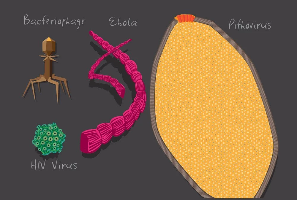
    giant viruses
    The genomes of giant viruses are several times longer, and the viruses themselves are much bigger, than those of smaller viruses. © Julie McMahon

    The discovery of enormous viruses, which are just slightly larger than some bacteria, provides a possible explanation. Some amoeba-associated viruses, such as pandoraviruses and megaviruses, may grow to a diameter of 700 nm, making them about twice as large as chlamydia-related bacteria. Not only that, but their genome is just as large as that of the Klosneuvirus, which was found in 2017 at an Austrian sewage treatment facility. The genome of the norovirus (found around toilets), which causes genital herpes, is 1.54 million bases long, or almost three times as long as that of the bacterial pathogen Mycoplasma genitalium.

    What is contained in the DNA of these gigantic viruses, however, is much more peculiar. This is because viral DNA includes genes for protein biosynthesis, the process by which cells manufacture the components for their metabolism and reproduction, in addition to the instructions for the construction of new viruses. It follows that these viruses at least have the genetic instructions for a lot of this cellular apparatus.

    Viruses in the primordial soup

    How, therefore, do these genes get into the body? To be more precise, both of the following scenarios are theoretically possible: First, it’s possible that these and other viruses originated from regressive single-celled creatures. Because of this, it is likely that viruses and all creatures with cells have a common ancestor. Then, when the viral forerunners transitioned into obligatory cell parasites, they reversed their development to eliminate all the parts of the cell they no longer required.

    Alternatively, viruses’ basic architecture might be traced back to the origins of life. Many scientists have speculated for a long time that RNA, not DNA, served as the hereditary material in the earliest cell-like structures that formed in the “primordial soup.” This suggests that modern RNA viruses may have descended from ancient RNA viruses.

    Suppliers of new genes

    Nonetheless, viruses may have played a pivotal role in the latter stages of evolution. The reason for this is that many of them can integrate their DNA into the host cell’s DNA. Occasionally, important sequences from these viruses are passed on to the next generation. Protein structures of viral origin have recently been detected in practically all domains of life, from bacteria to eukaryotic plants and animals.

    Our genome also contains viral gene sequences; in 2001, when the first human genome was decoded, researchers were shocked to discover 600,000 base sequences in our genome that were definitely of viral origin. Approximately 43% of our overall genetic material is now known to be composed of such viral DNA sequences, and some of these sequences have been shown to serve essential roles. Placental development, hormone generation in the adrenal glands and ovaries, protection against cancer, and proper muscle and brain function are just a few examples of how viral genes play an important role in the body.

    We should thus see viruses as a source of novel genes for cellular organisms, and not only as pathogens. Scientists think that viruses have helped cellular organisms evolve by transferring genes to them at various points throughout time. Simultaneously, some of these viral sequences may possibly be older than the period when viruses and cells had a common ancestor.

    Facilitator of evolutionary change

    Indirectly, viruses also pushed evolution forward by selecting novel features. Ten thousand mammalian proteins were compared for their rates of evolution in 2016. Therefore, it was clear that the rate of change for proteins involved in virus defense or other kinds of viral interaction has been three times that of most other proteins.

    Viruses seem to be responsible for around 30% of all amino acid modifications in the primordial section of the human proteome. This is the first proof that viruses play such a significant role in shaping adaptability. In spite of this, researchers find that encounters with viral infections mold not just our immune responses but also millions of additional proteins with seemingly no link to viruses or the immune system.

    Viruses pose a serious danger to human health, yet without them, the quality of life on Earth would likely suffer.


    Bibliography

    1. Nationalgeographic.com, 2020, “There are more viruses than stars in the universe. Why do only some infect us?“
    2. “Virus Taxonomy: 2021 Release”. talk.ictvonline.org. International Committee on Taxonomy of Viruses.
    3. Breitbart M, Rohwer F (June 2005). “Here a virus, there a virus, everywhere the same virus?”. Trends in Microbiology. 13 (6): 278–84. doi:10.1016/j.tim.2005.04.003.
    4. Koonin EV, Senkevich TG, Dolja VV, 2006. “The ancient Virus World and evolution of cells”. Biology Direct. 1 (1): 29. doi:10.1186/1745-6150-1-29.
    5. Lawrence CM, Menon S, Eilers BJ, Bothner B, Khayat R, Douglas T, Young MJ (May 2009). “Structural and functional studies of archaeal viruses”. The Journal of Biological Chemistry. 284 (19): 12599–603
  • History of smallpox: Vaccine, eradication, and the blankets

    Smallpox ravaged all humanity, starting in the years before Christ, reaching Egyptian merchants, North Africa, and India, and then to the most remote communities in the world through trade and conquests. The facial scars from this disease were even found on Egyptian mummies dating back to 1600 BC. The oldest smallpox outbreak case ever recorded happened in the war between the Hittites and Egyptians in 1350 BC.

    Treatment of smallpox, and the blankets

    It is known that the disease killed all of the civilization’s heirs, including the Hittite king and his son. The Hittite civilization is the first of many civilizations that were destroyed due to this disease. By 180 AD, the death toll from smallpox was 7 million. This caused a decline in the Roman Empire. The Spaniards and Portuguese carried this disease to the American continent, thereby breaking the Aztec and Inca civilizations. In the 100 years since the Spanish arrived in Mexico in 1518, the local population, which is thought to be around 22 million, dropped to 2 million.

    Millions of Indians were killed by the British and Americans, and half a million deaths were due to smallpox.
    Millions of Indians were killed by the British and Americans, and half a million deaths were due to smallpox.

    Along with colonialism, the whole world started to experience similar cases. In 1763, Jeffrey Amherst, the commander of the British armies in North America, was the first to unleash biological warfare by proposing to use the liquid obtained from scabs and blisters to apply it on blankets and give them away to the Indian tribes. In the 19th century, the American government used the same biological weapon against the Indians. Thus, smallpox caused the deaths of about half a million people, more than the number of people who died from wars and other epidemics before the 20th century. This is the most painful and neglected disaster regarding smallpox disease.

    Following Edward Jenner’s proven vaccination technique, this practice has spread to many parts of the world. Subsequently, in the 19th century, the cowpox virus was replaced by the vaccine virus, which was the first example of a vaccine obtained from cows. When administered orally or by injection to a person, the vaccine neutralizes or destroys offending microorganisms by causing lymphocytes to produce certain antibody proteins. The vaccine consisted of living, attenuated, or dead microorganisms (sometimes part of the protein that makes up the organism).

    World Health Organization to fight smallpox

    Dr. Donald Henderson was the head of the World Health Organization project.
    Dr. Donald Henderson was the head of the World Health Organization project.

    In 1926, the World Health Organization’s (WHO) Smallpox and International Vaccination Commission began to examine the content of the smallpox vaccines and set the production, storage, and distribution of the vaccines to a certain standard. Due to this inspection, vaccine production was brought under the control of Europe and North America until the mid-1900s. However, there was uncontrolled production in many parts of the world. Immunoglobulin, a vaccine produced by the human body against antigens and prepared by plasma in the blood of people who have recently been vaccinated, was released in the early 1950s. During a vaccination, this substance is used when the body can’t make enough antibodies on its own. Antibodies are the body’s defense against antigens.

    To summarize the treatment of smallpox, the smallpox vaccine was produced to a certain standard, and the introduction of the vaccine immunoglobulin produced by the human body resulted in the virus being removed from all of Europe and North America. This resulted in a worldwide effort to eradicate smallpox. The need for this universal effort and sensitivity comes from the fact that the variola virus causing smallpox only survives in a human cell, and if enough people get the vaccination, this means complete extinction of the virus.

    An international call from the Russian professor

    Zhadanov was the most important inspiration behind the elimination of smallpox in the 20th century.
    Zhadanov was the most important inspiration behind the elimination of smallpox in the 20th century.

    It was the Russian professor Viktor Zhadanov from the Soviet Union who proposed the need for a global program to eradicate the disease and led the treatment efforts beginning in 1958. At that time, the Soviet Union was a country with a vast territory where smallpox was common. At the same time, the WHO’s regional program aimed at eradicating smallpox on the entire American continent was the first joint international program implemented. The 12th World Health Congress, convened in May 1959, made the announcement of the undertaking of a universal effort to put an end to smallpox. This initiative was previously launched on the entire American continent in 1950 but began to weaken. At the 19th World Health Congress held in 1966, it was decided to use WHO funds for this universal effort.

    Even after this promising progress, the reluctance to continue the program still existed, and many people began to believe that it would not be possible to completely eliminate the smallpox disease. The suspicions were heightened by the fact that the remote settlements’ unknown situation, a lack of health services, logistical issues, and failures to eradicate malaria all contributed to the suspicions. However, the introduction of effective dry vaccines and the realization that this virus can live only in the human cell provided a great advantage in the struggle to root out the disease. 

    The last smallpox case in Somalia

    The last smallpox case was seen in 1977.
    The last smallpox case was seen in 1977.

    With the help of mass vaccination campaigns held between 1967 and 1980 and monitoring programs aimed at researching and detecting the types of smallpox throughout the world, the future was brighter. The Soviet Union contributed to the campaign with 14 million doses of vaccines and the United States with 190 million doses of vaccines. The last case of smallpox in the world was reported on October 26, 1977, in Merca, Somalia.

    To date, there are only two laboratories left that investigate the smallpox virus. One of these is the Moscow Research Institute in Russia, and the other is the Centers for Disease Control and Prevention (CDC) in Atlanta, the capital of the state of Georgia, USA. Even though the genetic sequence of the smallpox virus is known, there has been debate about whether the viruses from this latest case, which happened many years after the last one, should be kept to study or thrown away. 

    The world population is now too vulnerable to the possibility of the outbreak of new smallpox due to vaccination programs stopping after the disease has been rooted. There is growing anxiety about the possibility that this virus might be stored and used as a biological weapon by terrorists or governments. The complete destruction of the smallpox virus was postponed from June 1999 to June 2002.