What is a cataract? How was it discovered? Let’s talk about the history of thousands of years of retinal surgery. Blindness has been one of the biggest impediments that incapacitates humans. A person who loses his eyesight becomes constantly dependent on others to maintain his presence in society and even survive. All the developments made in improving eyesight can be considered among the most important scientific achievements.
Cataracts and diabetic retinopathy
The first studies began in about 1200 BC when the Chinese and Europeans developed the first glasses. Benjamin Franklin and many other scientists made numerous contributions to this field with their new invention of glasses; however, in the field of application, lenses were only made available to the public towards the end of the 19th century. The biggest inventions, such as contact lenses, which are constantly worn outside the cornea and improve eyesight, began to take place after that period. All these inventions were methods that could not prevent blindness but only strengthen eyesight. In this section, we will focus on cataracts and diabetic retinopathy, the two most common and treatable eye diseases.
History of cataract surgery
Cataract surgery is one of the safest and most effective surgeries and is performed on only more than 1.5 million people a year in the United States. The entire duration of the operation takes less than ten minutes today, and the patient can return to his normal life immediately. But cataract was not a disease that could be treated so easily in the past, and even then, there wasn’t a cure. The methods used to relieve the symptoms of the disease included nothing but pain and risk. Although more than half of people over the age of 60 have cataracts, the methods of treating this disease were first introduced only in the 1980s.
What is a cataract?
A cataract develops as a result of the transparent lens becoming cloudy, which is located behind the iris and focuses the light on the retina to create a sharp image. This inner lens consists of water and protein and transmits light. As the eye ages, unfortunately, some of the protein starts to pile up and fog the lens, affecting its ability to see. Since the cataract covers a large part of the lens, the lens must be removed surgically.

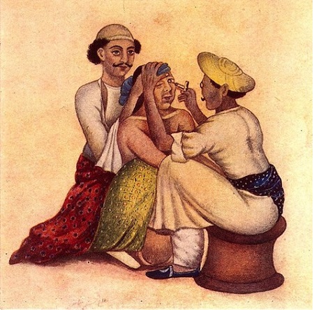
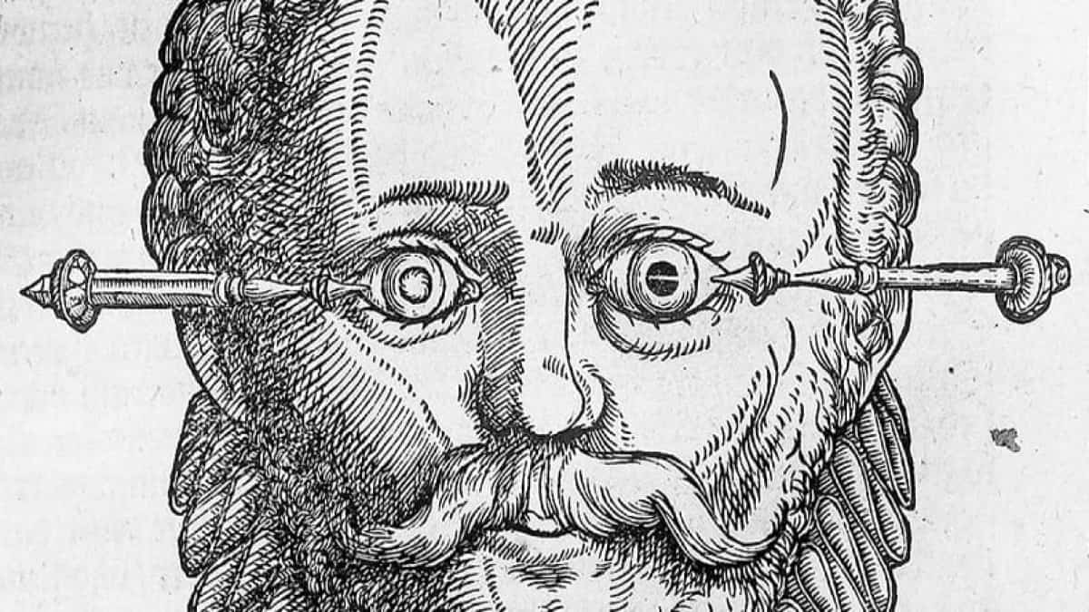
Ancient Egyptians used the “couching” method to treat this disease. In this method, the physician tried to rotate the lens that has lost its transparency by pushing it into the space behind it.
The coaching method was used for 1,500 years until 1748 when Dr. Jacques Daviel removed a patient’s cataract surgically. Unfortunately, since there was no anesthesia at that time, the patient whose cataract was removed would die from the pain. Towards the end of the 19th century, a new era for ophthalmology began with the use of cocaine as a local anesthetic. During these periods, cataracts could be removed through surgery, but after the operation, the patients had to stand still for two weeks with two sandbags between their heads. Also, after the inner cataract lens was taken out of the eye, the patient had to wear very thick-lensed glasses.
Fighter pilots and the invention of the intraocular lens
In 1949, everything in the field of eye surgery began to change in a good direction after Dr. Harold Ridley examined the fighter pilots’ eyes in World War II. These pilots were suffering from the tiny plastic pieces in their eyes coming from the destroyed windshield of the aircraft. However, this condition was not causing any side effects in any of the pilots. Based on this extraordinary situation, Ridley invented the world’s first intraocular lens using the same plastic material stuck in the pilots’ eyes. However, what he wanted to do was to remove the cataract lens and replace it with a transparent lens made of plastic. The physician community rejected this proposal from the very beginning. But the introduction of microscopes into surgical practice in the 1970s showed that Ridley was right on this point. Artificial intraocular lenses used by ophthalmologists today are quite flexible, foldable, can sit in a place with a very small incision, and provide better vision for millions of people.
Diabetic retinopathy
Apart from cataracts, other eye diseases cause blindness that can be treated today. Although cataracts remain the most common eye disease among older people, diabetic retinopathy is one of the common diseases that cause blindness in people under the age of 65 in the United States and many other countries. It is estimated that over fourteen million people in the US are diabetic. About half of these people are unaware of the illness that has not yet been diagnosed.
Diabetic retinopathy is a complication of diabetes caused by damage to the blood vessels in the retina. Damaged blood vessels in the early stages of diabetes cause blood and fluids to leak out of the veins and flow into the retina. This leak causes the sensitive nerve tissues in the retina to swell, letting the retina send distorted shapes to the brain and blur vision. As the damage in the blood vessels grows, some vessels supplying the retina occlude, and this prevents the necessary oxygen and nutrients from reaching the retina.
Diabetic retinopathy can be laser-treated. In laser therapy, the physician sends energetic laser light to the retina layer of the eye to close some parts of the retina, which have lost their function because of leaking abnormal vessels or blood loss. This treatment method has preserved the eyesight of millions of people who would have gone blind.


