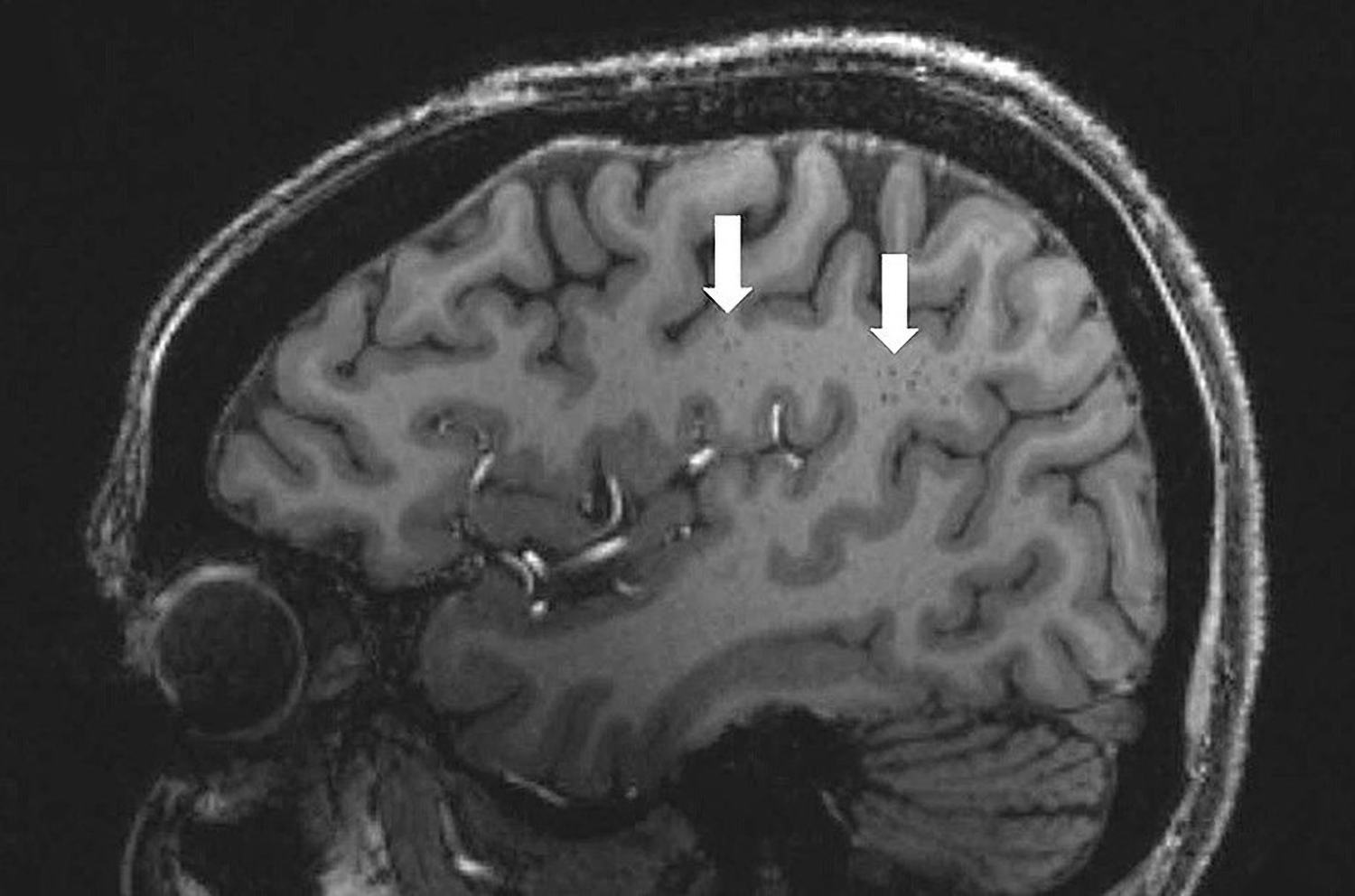
High-resolution brain scans have recently shown that migraine not only presents itself in the form of frequent headache episodes but also shows up on the brain. There are permanent alterations. The results indicate that those who suffer from migraines tend to have noticeably larger cavities surrounding the blood vessels in certain brain areas. They also tend to have more microlesions, or minute holes, in their blood vessels. Those who suffer from migraines may have a neural lymphatic system malfunction, according to the findings of the study.
Mild darkening of the brain’s central white matter is indicative of abnormally enlarged fluid-filled gaps surrounding blood vessels, as shown in this migraine sufferer above.
Migraines cause severe headaches, sensitivity to light, and nausea for around 15% of the world’s population (1.1 billion people) and around 11.4% in the US. We still don’t fully understand what sets off this illness, which has a weak hereditary component, or how it shows itself in the brain. But it does seem that migraines have other, non-headache-related manifestations. Between headache episodes, migraineurs exhibit anomalies in brain activity, cerebral cortex structure, and specific membrane lipids in the blood.
A picture of a brain with migraine
An additional characteristic of migraines has been found by a team led by Wilson Xu of researchers at the University of Southern California in Los Angeles. They scanned the brains of 25 patients: 10 people with chronic migraine, 10 people with episodic migraines, and 5 healthy control participants. The researchers doing this MRI scan were especially interested in the minuscule alterations near the brain’s vascular system.
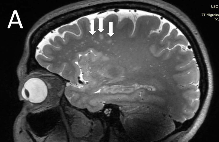
This is the first research to our knowledge to employ ultra-high-resolution MRI images to investigate microvascular alterations in the brains of migraine sufferers. Pain during a migraine episode has long been thought to be caused by a disruption in blood flow to the brain. Now, Xu and his team sought to see whether these shifts could be detected outside of the context of sudden strikes.
A look into the brain’s blood vessels
The group did, in fact, uncover its target. Migraine sufferers, both those who experience them often and seldom, tend to have alterations in the perivascular spaces. These are lymphatic drainage channels located around the brain’s blood vessels. The presence of degenerative changes or inflammation of the vessels is sometimes accompanied by the symptom of dilated blood vessels.
The findings showed that the perivascular spaces were most enlarged in the so-called centrum semiovale in the migraine sufferers. White matter, which consists mostly of nerve conduits, is primarily located in this area of the brain between the cerebral cortex and the central ventricles. This crescent-shaped region of the brain is located on both sides, and the researchers detected an increased number of microlesions and other microscopic, concentrated regions there due to tiny breaches in the brain’s blood vessels.
The brain’s waste disposal system
Migraines, as Xu and his colleagues see it, may be related to an underlying dysfunction in the glymphatic system, the network of tubules, cavities, and drains responsible for removing waste from the brain. The perivascular spaces contribute to the brain’s waste management system. More research on their role in the development of migraine might aid in understanding the disorder’s complicated causes.
However, it is still unknown if the current alterations seen are a result of migraines or the cause. Researchers are hoping to learn more about this by conducting bigger investigations over longer periods of time.


