Anatomy of Heart in Ancient Egypt

Cardiology in ancient Egypt was practiced by physicians employing an art understood according to the possibilities of the time and indicated in medical papyri.
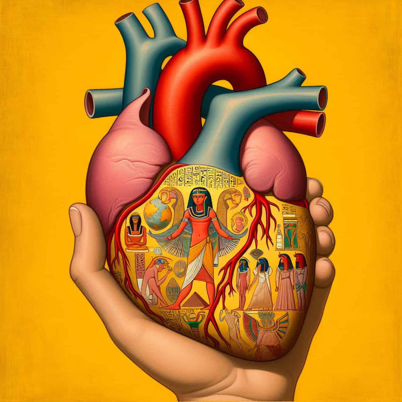
Anatomy of Heart in Ancient Egypt

Cardiology in ancient Egypt was practiced by physicians employing an art understood according to the possibilities of the time and indicated in medical papyri. This medicine was daily practiced by religious or civilian physicians already well-versed in the anatomical, physiological, and clinical knowledge of that era. The practice was also conditioned by the available pharmacopoeia.

- Advertisement -

The Heart

The heart is an organ depicted and perceived in several distinct ways in Egypt.

Lexicography

There are several lexical designations for naming the heart in ancient Egyptian. Normally, only the first two, “jb” and “ḥ3ty,” are encountered in medical texts. The term “nṯry” can be used in a more religious context.

The “jb” Heart (ib)

image
heart jb (ib)

The “jb” heart can refer to internal parts of the body, as in “the heart of a tree” and the “innermost being.” In the latter case, “jb” corresponds to “the intellectual and emotional organ, both conscience and thought; it is also the memory of the being.”

The “ḥ3ty” Heart (haty)

image 1
heart-ḥ3ty

ḥ3ty, “the heart,” the “actual cardiac muscle,” and also “as the seat of feeling” (in butchery scenes), “Herzmuskel” (cardiac muscle); “the heart,” and also the seat of feelings, dem. ḥ3.t(y) “heart.” Then, we find in Coptic the expression ϩⲏⲧ SB “heart” as a body part, and also “sense, intelligence,” ϩⲉϯ F “heart.”

The “nṯry” Heart (netchery)

image 2
Heart-nṯry

nṯry, “the heart”. The Gate of Isis, Cairo, 1999, p. 34, 103, 185, in the phrase “rwj ḥ3-jb r nṯry n ḥm.k” “drive away the sadness (ḥ3-jb) from the heart (nṯry) of your majesty”; see also for the same meaning: Karnak, Temple of Opet, Roman era, Augustus (KIU 4238,9), B. Mathieu, “And all of this exactly according to his will.

There is a hymn in which it is said that “Re-Khnoum… fashioned… (nṯry ḥr sšm sm3 ḥr rmn) the heart organ for distribution, (and) the respiratory system for transport” (250,11b–250,12a–b). This idea of the human body is talked about in “Et in Ægypto et ad Ægyptum: Collection of studies dedicated to Jean-Claude Grenier,” CENIM, 5, Montpellier, 2012, pp. 506, 508, note (z) pp. 514, 516. These are precise anatomical and physiological notions situated in a religious context that have a fundamental effect: maintaining life through cardio-circulatory-respiratory action.

- Advertisement -

Egyptian Hieroglyphs

The iconic aspects of the heart in Egypt have varied since ancient times, then stabilized in the Old Kingdom into just a few forms with their derivatives to constitute a relatively stable group of signs until the Late Period. Regarding the paleography of the different variants of the Egyptian hieroglyph representing the heart (F34), after the simplest oval, two forms early on stabilized, one solidly cut, and the other presenting details in its internal portion.

The Unicode hieroglyphic font only has the solidly cut form:

A mammal’s heart

Regarding the identification of the hieroglyphic heart, its forms sometimes vary slightly, but enough to allow the observer to compare them to those of a bovine organ (wider), a sheep (longer), a pig (more regular), or even a human (seen in the tombs of Pepi II, Mereruka, Nefertari, etc.).

It is, in any case, the representation of a mammal’s heart.

- Advertisement -

A vase-shaped heart

Another solution would be to see in this sign a vase with two handles and a border at the neck level, which could be explained since butchered hearts were sometimes placed in comparable pots as well as in wider containers without handles. For example, in Esna, one of the signs takes the form of a granite vase ȝb (W7), knowing that the name of the city of Elephantine can be written using this vase as “containing a heart.” It should also be noted that representations of heart-shaped vases were used to represent the heart in the New Kingdom.

It should also be noted that sometimes the tracing of the sign hesitates between rendering a pot in its upper portion, with however internal details specific to a visceral interpretation (see in the tombs TT414, of Ânkh-Hor).

Here, symbolism is expressed, not always a real anatomical diagram, as with the problem of the Egyptian “opposite hand.”

Values

Classically: logogram jb “heart”, determinative of ḥty “heart”. More rarely, the ideogram “heart”.

- Advertisement -

Variants

Late variants with lateral details

Some late variants with lateral details: F132, F132A, F132B, and F132C (they are not included in the Unicode font).

The compound sign

The heart is also part of the sign nfr (F35), variant F35A, and other variants (they are not included in the Unicode font). With none (F35A), one, or two perpendicular bars placed on the upper part of its rising branch. This sign has been fairly stable since the Thinite period.

The heart is here drawn with details in its internal portion.

- Advertisement -

Anatomical Pieces

It is clear that all of the drawings of the heart in Egyptian hieroglyphics came from looking at real body parts that had been cut out, usually during butchery dissections. Indeed, we find specimens of these in scenes of butchery and, subsequently, on offering tables.

Two hieroglyphic forms early on stabilized, one solidly cut and the other presenting details in its internal portion. When these details are visible, it is the right atrium that is, in the best cardiac reproductions, opened at the top, that is, cut. In hieroglyphs of very good quality, this cutting action reveals an internally vertically pectinate (Mereruka’s Tomb) or crosshatched (Tombs of Pepi II; Senusret I; Ramses II, etc.) portion, or sometimes, painted white for better visibility (Nefertari’s Tomb). Then, for the lower part, it is now the left ventricle that is most exposed at the bottom and will also be presented open (cut) and pectinate (fleshy columns).

Protrusions are shown emanating from the heart on both opposite sides and/or at its upper part. By looking closely at the earliest drawings from the Thinite period, it is clear that all of these protrusions first stood for long blood vessels that were attached to the organ and leaving it. These vessels were cut short enough to make the writing mark. Six large vessels with the aorta, pulmonary trunk, and the four pulmonary veins, plus the two vena cavae, seem to be depicted in representations (Tomb TT 100 of Rekhmire; a figure from the Cairo Museum) or even in an exceptional engraving such as that of Adjib (Am. N.F. I 33, Thinite Period), which additionally adds two coronary vascular processes adhering to the myocardium. The other representations seem more random.

Along with these free hearts that look like hieroglyphs or almost hieroglyphs and were taken from animals as offerings, there are also more realistic images that show a desire to get closer to the truth of anatomy.

- Advertisement -

For example, some of these representations show a central depression vertically dividing the myocardium into two, housing a large conduit. This should show the two hearts: the right and the left. Thus, we find partitive notions in representations of several anatomical offering pieces (Nebamon, British Museum EA37985; TT 38 of Djeserkareseneb…).

For example, in the group of free forms representing the heart, some particular variants appear due to their superior and lateral graphic additions (TT 277 of Ameneminet; Pyramid of Tanoutamon; LdM of Hor; Louvre N 3209; Tomb QV 66 of Nefertari). Like in the corresponding late hieroglyphs, these units have formations topping them and/or in the shape of two sharp wings, which often protrude upwards and outwards, placed on each side (F 132 A, F 132 B, and F 132 C, which are not included in the Unicode font). This is the pericardium.

It should also be noted that the fatty-serous apparatus of the heart, with the fatty fringes (ḏ) of the pericardium clearly visible in an open mediastinal situation, is described in medical texts (e.g., Ebers papyrus 855, n. 101, 13c–14), which we will address regarding pathology, where this fat is accused of causing cardiac disorders.

Heart Anatomy through Amulets

Amulet of the Heart (Egyptian Museum of Turin, inv. C.1192).

Heart-shaped amulets in ancient Egypt were made from all the usual materials used to make these small, precious or non-precious objects. Although quite often similar to the corresponding hieroglyphic forms, amulets representing a heart sometimes offer additional interesting details.

- Advertisement -

For example, one of them (Louvre E 2159) represents a heart laden with part of the remainder of its pericardial mantle. Then, its upper part is crossed by two horizontal lines separating the atrial part and the ventricular part.

On other of these heart-shaped amulets, the beginnings correspond to the simplified start of a Y-shaped (British Museum EA14705) or T-shaped (MET 26.8.144; Louvre AF2333; Chicago OIM E21296E…) engraving, which also delimits a double atrial part at the top and two lateral ventricular parts left and right at the bottom. The vertical middle part corresponds to the interventricular septum. The groove separating the left heart from the right heart is still visible in late amulets (two bas-reliefs of Ihy, from the Temple of Dendera).

Among the compound forms, some show a lower wound with crosshatching (Louvre E 14708; Louvre E 22620; British Museum EA18284; British Museum EA23427, Turin inv. C.1192: photo opposite…), reminiscent of the fleshy columns already mentioned above.

The large conduits of the heart in anatomical context

Located within the vital body envelope, which is vascularized from its sšmt “conducting system” (Berlin Papyrus 3038 163 b, 15.5) by certain hollow mtw channels (vessels) related to the different jb when they designate internal parts, the true heart ḥȝty is considered by medical texts as having its “normal place” (st) “at the center of the pulmonary tract” (Ebers Papyrus 855 k. 101, 5-8), and its apex is clearly perceived to the left (Ebers Papyrus 855 n. 101, 12).

- Advertisement -

Located in the anterior and middle mediastinum, the “mediastinal viscera,” called bskw, are composed of the remnants of the thymus called sḫn in front and above, then the interweaving of various large conduits mtw (vascular, airway, digestive, nervous, lymphatic), and the heart.

Located in the posterior mediastinum, we find the esophagus behind and the trachea-artery in front. They can be called ḥtyt (Urk. IV, 482, 12)  and šbb (Smith Papyrus 28. 9, 19).

A very important vessel mt and conducting a fluid (mw) is called šspw “the receiver” (Ebers Papyrus 855 c. 99, 19). The aorta (a large empty mt) gradually passes behind the esophagus. The mtw vessels are supposed to transport blood, water, and air. After death, the collapsed arteries seem filled with air, while the veins contain coagulated blood, which will lyse and subsequently liquefy.

Here is what a passage from a major medical papyrus, papyrus Ebers 856 b. 103,2c-3b, tells us: “As for the man, twelve vessels (mtw) are in him for his heart (ḥ3ty); they are the ones that give to each place of his body (t).” This text indicates that twelve vessels are connected to the heart, including the usual eight larger vessels, which are the superior vena cava, inferior vena cava, aorta, pulmonary trunk, and the four pulmonary veins, plus the coronary sinus, right coronary artery, left coronary artery continuing into the circumflex artery, and an interventricular artery. In another medical papyrus, a passage from the Berlin Papyrus 3038 (163 b. 15, 5-6) counts no less than twenty-two, and these conduits are said to establish themselves between the head and the heart. These are the ramifications extending from the large arterial (or venous) vessels joining the head, e.g.:

- Advertisement -
  1. the aorta,
  2. the right brachiocephalic artery,
  3. the left common carotid artery, continuing with the carotid arteries
  4. right internal,
  5. right external,
  6. left internal,
  7. left external, the brachiocephalic veins
  8. right
  9. left continuing with the jugular veins
  10. right internal,
  11. left internal
  12. right external,
  13. left external, etc.

plus a central thyroid, eight and fourteen make twenty-two.

Anatomical Conclusion

Heart amulets MET DP109375 1 1
Heart Amulets (MET 10.130.1702 & 10.130.1804)

For pure anatomy, one must resort to the various forms taken by the sign, each expressing a complementary perspective for this asymmetrical organ and therefore more complex to render in art. After synthesis, the Egyptian physicians knew: the myocardium, the two atria, the two ventricles, and some of their internal structures due to their irregular shapes (fleshy columns), as we have seen. Then externally, being clearly visible, naturalists had seen the eight large vessels connected to the heart, which are (here designated by our modern denominations): the superior vena cava, inferior vena cava, aortic arch, pulmonary trunk, the four pulmonary veins, plus coronary arteries and veins. Both butchers and dissectors saw the pleura extending beyond, as well as the pericardial sheets.

All of this is compatible with macroscopic observation.